Case of the Week: December 18, 2019

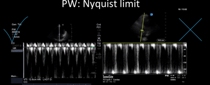
This is a 70yo F with a recent diagnosis of left sided lung cancer. She presented with hemoptysis and was subsequently admitted to the ICU for monitoring.  There she was found to be tachycardic at 120bpm and had reduced urine output. The patient had no known cardiac history and no previous echocardiogram on file. The POCUS team performed a focused echocardiogram, given that she was a newly admitted patient with ongoing tachycardia. 

By |2019-12-19T03:22:55+00:00December 19th, 2019|Case of the Week, Cases|Comments Off on Case of the Week: December 18, 2019

Case of the Week: December 9, 2019

This week's case is a 67-year old woman who presented for an outpatient EGD. Post-procedurally she became hypoxic, so was brought to the ICU for monitoring and NIPPV. She was initially hemodynamically stable; however, over the next 6 hours she became profoundly hypotensive requiring very high doses of multiple vasoactive agents. She had a PMHx significant for severe COPD and HFpEF, with an Echo from earlier this year showing an EF of 50%.Below are her the clips taken upon arrival of the POCUS team the next morning. What do you think is causing her shock based on these images? You might want to make yourself comfortable, cause this week's case is a doozy and has a lot to unpack.*Note: given her marked hyperinflation she had no obtainable traditional parasternal or apical windows, so all views were obtained from a modified subxiphoid position (hence the unconventional axis). These scans are a good lesson, however, that you can still get lots of important information even when your views aren't perfect!

By |2020-09-19T05:51:00+00:00December 9th, 2019|Case of the Week, Cases|1 Comment

Case of the Week: December 4, 2019

85 yo M polytrauma from a high-speed MVC. Now recovering in the ICU, still intubated, but over the last few days has had worsening oliguric AKI and is now on 10mcg/min of NE. The POCUS team was asked to perform a “volume status” assessment. This is one of the most common requests we get. Below is a representative look at a typical set of images we try to acquire to help answer the million-dollar question of whether a patient would benefit from fluids. Have a look at the images below (which, be forewarned, are quite Doppler heavy) and see what you think!

By |2019-12-06T22:27:22+00:00December 4th, 2019|Case of the Week, Cases|Comments Off on Case of the Week: December 4, 2019

Case of the Week: November 20, 2019

This is a case of a 52 yo F trauma patient with severe traumatic brain injury. She was intubated and transferred to the ICU where, given the severity of her TBI, an intraparenchymal ICP monitor was placed by neurosurgery. The ICP monitor was initially showing an ICP of 10. Overnight, however, despite no apparent clinical change, the ICP waveform was inconsistent and the readings were now showing values around 40-50. We thus performed a point-of-care TCD to help determine whether her ICP was truly elevated or whether the ICP monitor was giving spurious information. Have a look at the following images. Do you think the TCD spectral waveform is consistent with markedly elevated ICP?

By |2019-11-20T18:24:02+00:00November 20th, 2019|Case of the Week, Cases|Comments Off on Case of the Week: November 20, 2019

Case of the Week: November 11, 2019

This is a 40-year old man status post-liver transplant (several months prior) who has had a complicated hospital course. He is now re-admitted with presumed septic shock (query HAP) and has been intubated for respiratory failure. He's also had a formal abdominal ultrasound commenting on periportal hepatic edema and signs of congestion.Based on the images below what interventions might you make to the team in addition to antimicrobial therapy? The team is particularly worried about right heart failure and elevated right-sided pressures - what do you tell them based on your findings?

By |2019-11-11T17:45:58+00:00November 11th, 2019|Case of the Week, Cases|Comments Off on Case of the Week: November 11, 2019

Case of the Week: November 7, 2019

This is a 54 yo M who presented with acute hypoxic respiratory failure necessitating intubation and ICU admission. Post-intubation he had a P/F ratio of 54 despite a relatively unremarkable CXR. The PEEP was increased to try to improve oxygenation. A CTPA was ordered which was negative for PE and showed just a small right lower lobe consolidation. Given that the refractory hypoxia seemed discordant with the pulmonary pathology, concern was raised for an intracardiac shunt. A point-of-care TEE was performed including a bubble study. Have a look at the selected clips below and see what you think!

By |2019-11-07T14:57:11+00:00November 7th, 2019|Case of the Week, Cases|1 Comment

Case of the Week: October 28, 2019

This is a 74-year old gentleman with multiple medical comorbidities who was admitted to the ICU with septic shock and bacteremia. He's had a tumultuous course in the unit and has been difficult to wean from vasoactive agents. Below are two series of images. The first were taken earlier in his stay, while on higher doses of vasoactive agents and more acutely unwell. The second were taken a week later when he was clinically significantly improved and weaned from inotropic support; however, there was concerned that some of his Echo parameters had actually worsened! What are we looking at here, and what's the explanation?

By |2019-10-28T09:28:50+00:00October 28th, 2019|Case of the Week, Cases|Comments Off on Case of the Week: October 28, 2019

Case of the Week: October 22, 2019

This is a case of a 56 yo F admitted with sepsis and MSSA bacteremia. A CT Head revealed several lesions concerning for septic emboli. She had known chronic significant mitral valve pathology documented prior to her admission. The POCUS team keen to see this pathology and to look for any additional signs of infective endocarditis. What do you notice about the valve on the 2D echo images and what specifically do you see on its colour doppler interrogation?

By |2020-06-07T21:13:53+00:00October 24th, 2019|Case of the Week, Cases|Comments Off on Case of the Week: October 22, 2019

Principles of Doppler

Principles of Doppler Learn the principles of Doppler ultrasound in this 20-minute screencast from Dr. Katie Wiskar. This tutorial covers basic ultrasound physics, different Doppler modes, and multiple examples from various POCUS applications.Relevant ScreencastsTutorial on aortic stenosis vs sclerosis: https://westernsono.ca/aortic-stenosis-vs-sclerosis/Tutorial on transcranial Doppler: https://westernsono.ca/screencasts/miscellaneous/trans-cranial-doppler-tcd/Tutorial on calculating stroke volume using LVOT VTI: https://westernsono.ca/screencasts/echo/stroke-volume-determination/

By |2020-04-22T22:49:38+00:00October 21st, 2019|Uncategorized|Comments Off on Principles of Doppler
Go to Top