Case of the Week: September 18, 2019

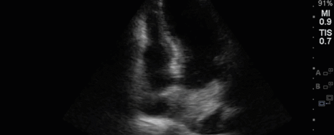
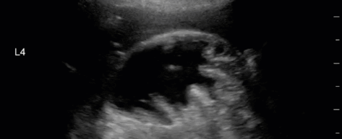
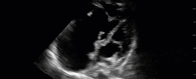
This week is a case of a 29 yo F with known severe pulmonary hypertension. The etiology was thought secondary to cocaine-induced idiopathic pulmonary arterial hypertension. Unfortunately, she sustained a cardiac arrest (you’ll see why when you look at the images). ROSC was obtained and she was transferred to the ICU. Despite maximal medical support of her RV (optimal ventilator management, IV flolan, inhaled nitric oxide, inotropes including milrinone and vasopressin) she had persistent hypotension and worsening renal failure necessitating CRRT. The overnight team decided to trial some small boluses of crystalloid to see if that would help. Have a look at the images below and decide whether or not you would give fluids or recommend something else? If I said the CVP (as measured from the right IJ central line) was 22, what would the estimated RVSP be?

By |2019-09-19T01:52:52+00:00September 18th, 2019|Case of the Week, Cases|1 Comment

Case of the Week: September 12th, 2019

It’s a 78 yo M with unwitnessed syncope, a subsequent tib-fib fracture, who was eventually admitted to the ICU for persistent hypotension and altered LOC that had not been fully elucidated. He had an extensive work up including a negative CTPA, CT head, and ultimately even an angiogram (based on some transient diffuse ST depression and a positive troponin) which showed clean coronary arteries. He eventually stabilized with good supportive care, and the ICU team was now trying to wean him off the ventilator and were aggressively diuresing him. They asked for a POCUS assessment to help guide further volume management. Have a look at the images. What two major findings are most striking? Should the team continue to diurese him or perhaps give some volume back?

By |2019-09-12T16:24:42+00:00September 12th, 2019|Case of the Week, Cases|Comments Off on Case of the Week: September 12th, 2019

Case of the Week: September 6, 2019

This is a 39 yo F post cardiac arrest NYD. She was in the weaning stages of her care and close to extubation, but she became newly febrile with increasing oxygen requirements. Her sputum culture was positive for E. coli. A portable CXR was done which did not show any obvious large consolidations. A POCUS thoracic study was performed. An unusual finding was seen on the left side (shown in the clips below). Also, to orient those who aren't familiar with the WesternSono shorthand here is a legend for the labels: L1 = Left anterior chest wall, L2 = Left anterior axillary line, L3 = Left costophrenic view, and L4 = Left PLAPS (PosteroLateral Alveolar and/or Pleural Syndrome)

By |2019-09-07T02:36:01+00:00September 7th, 2019|Case of the Week, Cases|Comments Off on Case of the Week: September 6, 2019
Go to Top