Answers to Review Questions & Extra Images 2
Answers
How can you manipulate your ultrasound image to better appreciate lung pulse?
Turn down the contrast, put the pleura at the focal zone, use a linear probe, view in M-mode, put the probe perpendicular to the pleura
True or False: Loss of lung sliding means a diagnosis of pneumothorax
False. Loss of lung sliding can also be caused by a mucus plug, pneumonectomy, mainstem intubation, atelectasis, therapeuedic pleurodesis, and emphysema blebs. If you are really keen on learning more, check out this video
What artefact is produced due to fluid or fibrosis?
B-lines!
True or False: B-lines rules out pneumothorax at its location.
True. The presence of B-lines indicated that the pleura are in contact with each other
You notice B-lines on your lung ultrasound image and see that the pleura is irregular and jagged. What is more likely, cardiogenic pulmonary edema or an inflammatory/fibrotic process?
Inflammatory/fibrotic process
You see a single B-line when putting your probe against a patient’s dependent zone. What is this likely to be?
This is likely normal lung tissue. <3 B-lines in the dependent region is generally normal.
What does the shred sign signify?
Non-translobar consolidation
True or False: Complex effusions have blood flow.
False. Consolidated lung has blood flow
How can pleural effusions be classified?
Small: 0-2.5 cm between lung and chest wall
Moderate: 2.5-5.0 cm
Large: >5cm
How do transudative and exudative effusions appear on ultrasound?
Transudative = black, exudative = hypoechoic (& less commonly isoechoic or hyperechoic)
Images
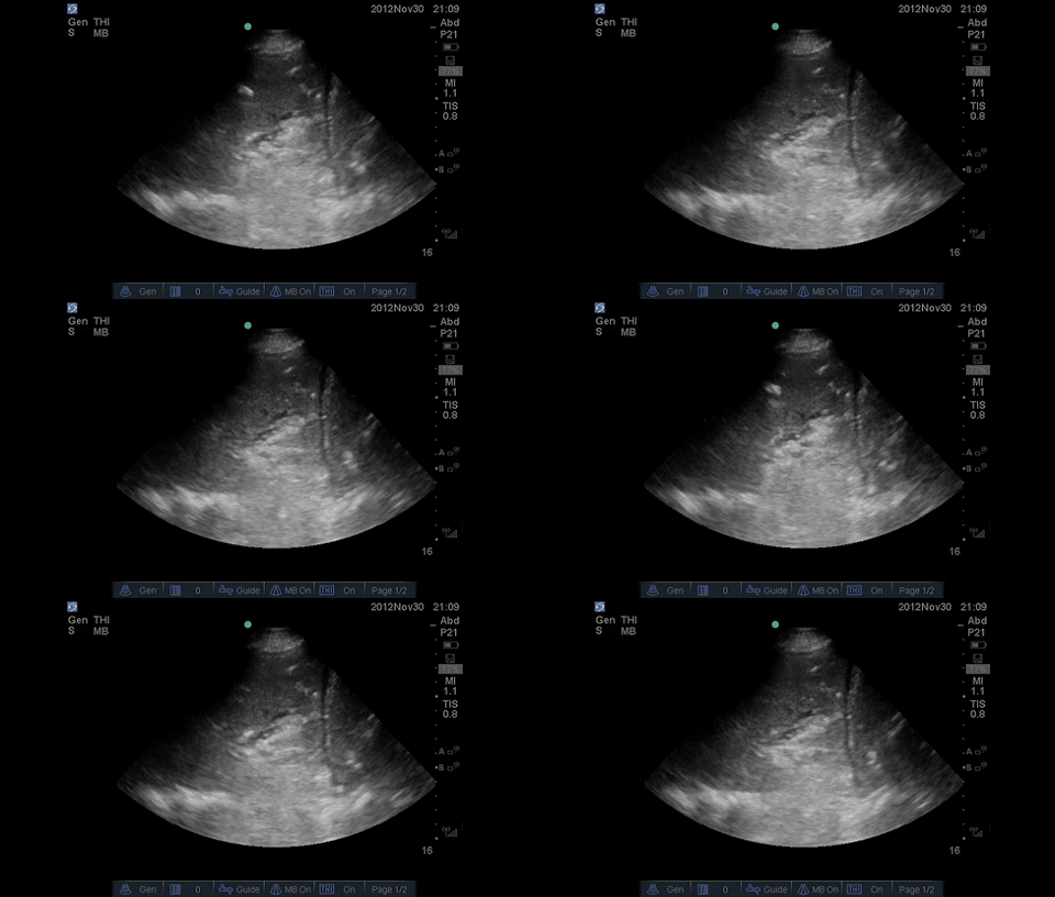 Here we have a dense consolidation with some dynamic air bronchograms
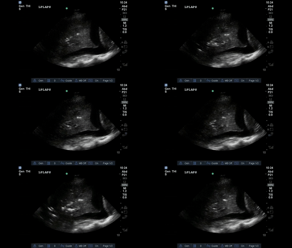 Some more beautiful dynamic air bronchograms with a pleural effusion
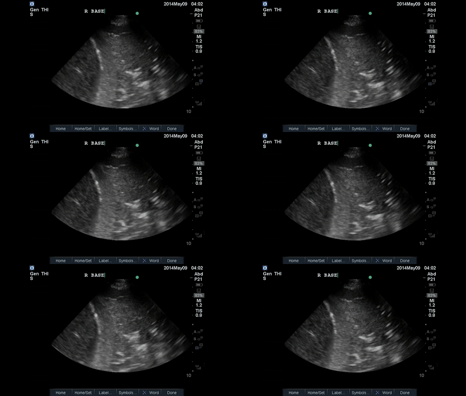 You are looking at hepatization with some pus
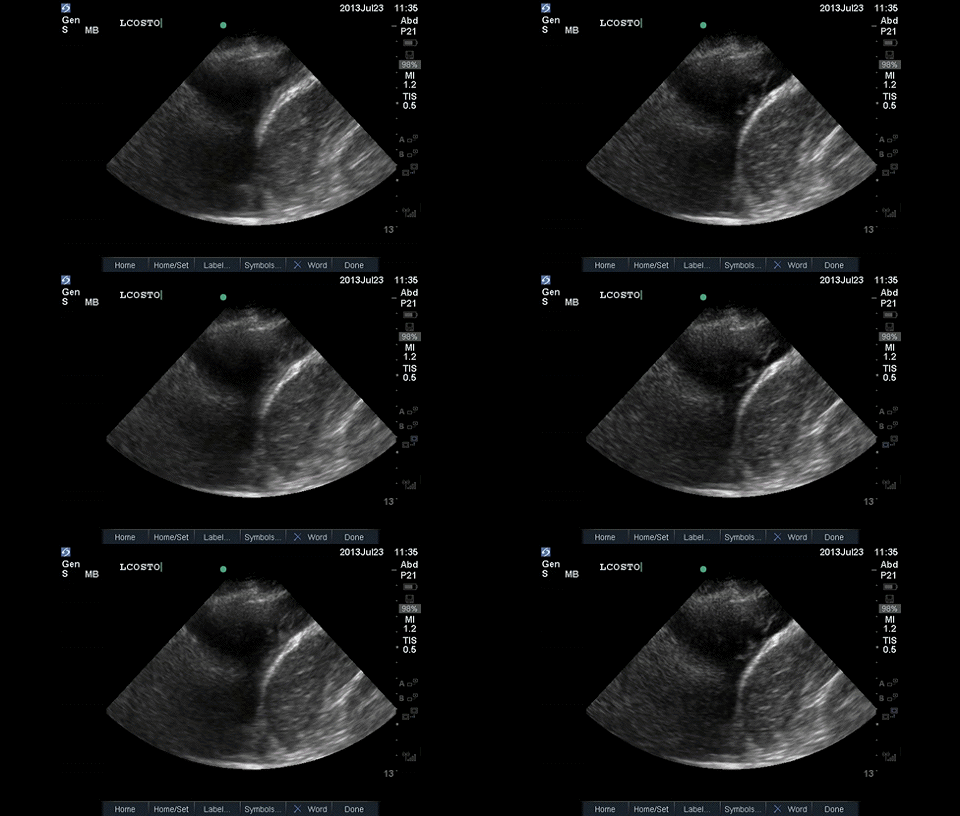 This is a complex pleural effusion
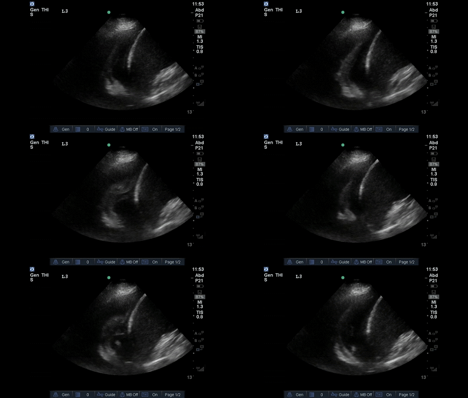 Pleural effusion with the flapping sign
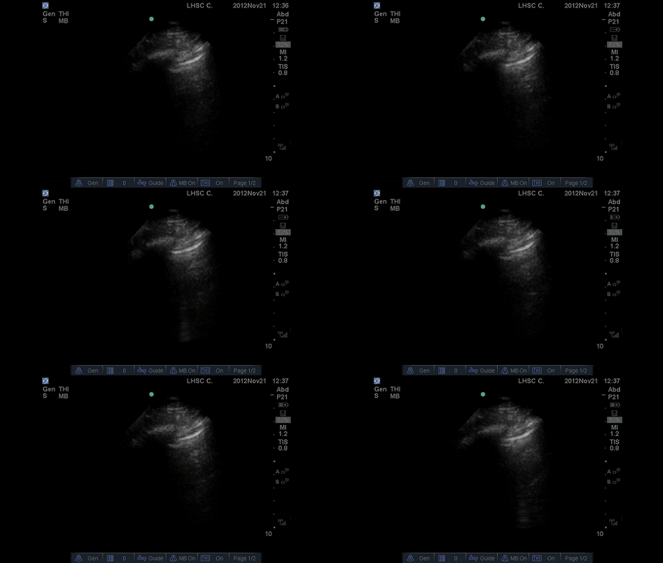 Can you see the lung point?
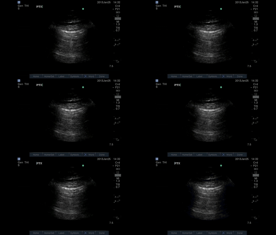 Here there is no lung sliding
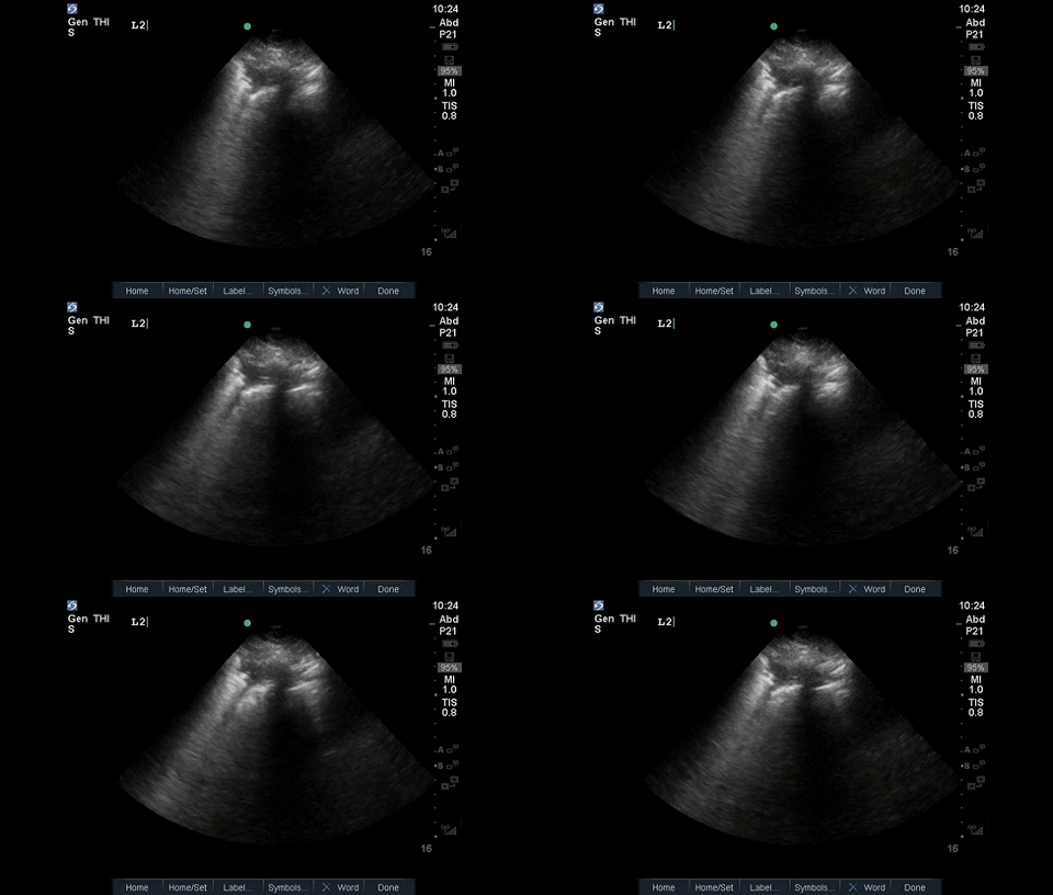 Finishing off with a shred sign