This annual course is offered as the official course of our Critical Care training program and is attended by all critical care fellows. Further, it will be the most comprehensive course offered in the country for the intensivist or resuscitative physician who seeks to acquired fundamental skills in the assessment of:
-Thoracic and lung ultrasound
-Vascular access, including peripheral veins and arteries
-Assessment for DVT
-Critical Care Echocardiography including assessment of LV function, RV, pericardium, valves and IVC.
The course will be directed by Robert Arntfield.
Additional faculty, agenda and brochure will be announced closer to the course but for a sense of what is in store for you, see the images from the 2014 course.
If you have interest in attending this course please email [email protected] who will be able to notify you when registration for this course opens early in 2015.
]]>Hosted at the state of the art Canadian Surgical Technologies & Advanced Robotics (CSTAR) facility, this two-day symposium provided medical students with an introduction to point of care ultrasound. The event was attended by 38 medical students from 6 medical schools across Canada. Extensive opportunity for “hands-on” skill development was a focus of the symposium. This practical component was enhanced by didactic teaching from local experts. The weekend was capped off with fierce competition and a showcase of ultrasound skills as participants attempted to take home the Western SONOGames Cup!
The first day began with an electrifying keynote address by Dr. Rob Arntfield. Dr. Chris Byrne, the founder of the 1st medical student ultrasound symposium, then gave participants an informative lecture highlighting the fundamentals of ultrasound. This was followed by exciting workshops on Cardiac and Lung ultrasound led by Dr. Rob Arntfield. Dr. Drew Thompson wrapped up a busy first day by leading the FAST and Obstetrical ultrasound workshops.
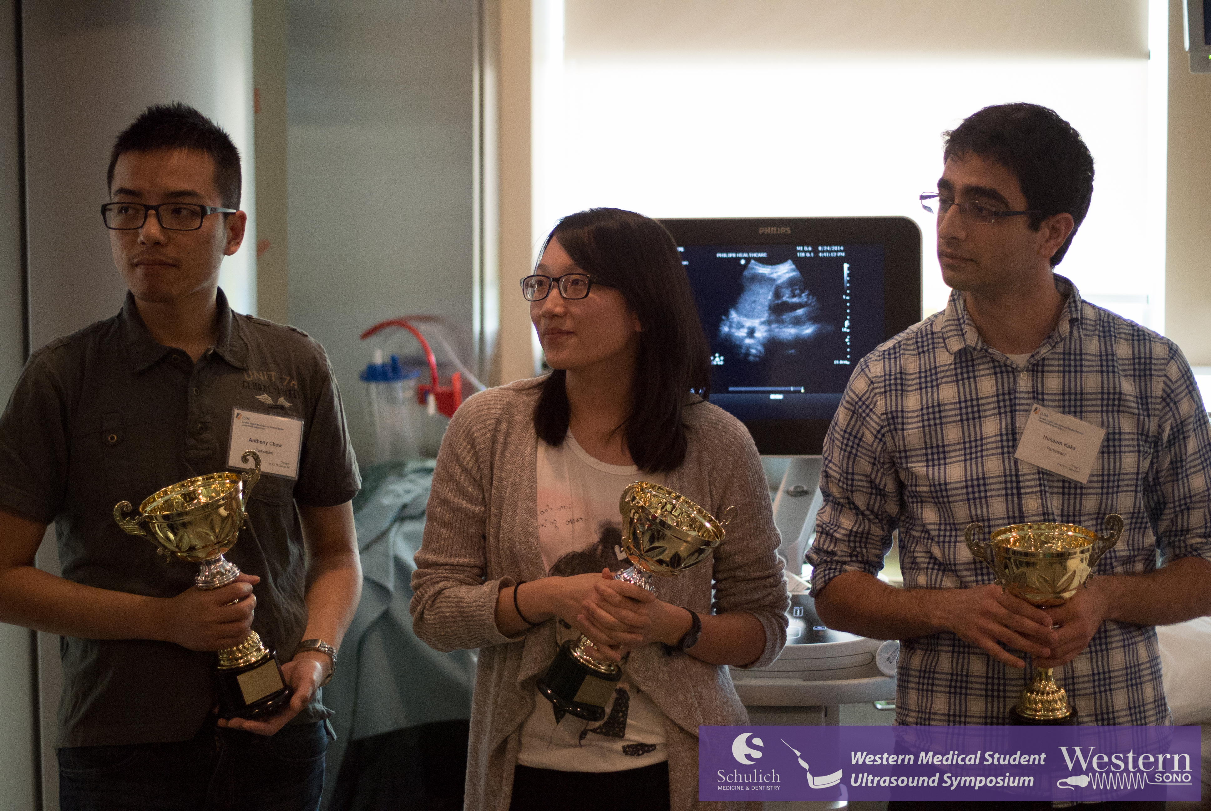
Day two began with an engaging lecture by Dr. Naveen Poonai highlighting the use of ultrasound in pediatric fracture localization and reduction. Dr. Drew Thompson then led instruction in Abdominal, Aorta and Hepatobilliary ultrasound. This was followed by an expanded session on ultrasound in Procedural Guidance, taught by Drs. Vishal Uppal, Jon Brookes and Rakesh Vijayashankar. The symposium concluded with fierce but friendly competition during our Western SONOGames, which included an expanded emergency medicine simulation station (led by Drs. Ouellette and Davis). Congratulations to this year’s SONOGames winners (see photo below) for clinching a close battle and taking home the hardware. The bragging rights are all yours!
We would like to extend a special thank-you to our corporate partners SonoSite Canada, Phillips Canada, and General Electric Canada for the donation of state-of-the-art ultrasound machines. The support of ultrasound machines and expertise was invaluable! We were also fortunate to have MedaPhor graciously provide a virtual-reality training system – ScanTrainer – for medical students to explore and hone their ultrasound skills. Finally, this weekend could also not have been a success without our standardized patient models who volunteered their time to support the event.
We are excited for the new academic year and what it will bring for PoCUS and undergraduate medical education. The experiences from this past weekend demonstrate that medicals students are ready to embrace point of care ultrasound and will seek it out additional instruction independently!
Below are some photos from the weekend with credit to Weiang Yan. Also find the members of the medical student planning committee whose tireless efforts cumulated in this weekend’s symposium and the faculty and residents whose enthusiasm and expertise in teaching made the weekend a success.
Thank you to everyone for your support as we look forward to 2015 and the making of the 3rd annual Medical Student Ultrasound Symposium!
Medical Student Planning Committee: Nick Packer (2015), David Mainprize (2015), Mike Clemente (2015), Weiang Yan (2015), Ryan McLarty (2015), Evan Ailon (2016), Josh Burley (2016), Justin Cottrell (2017), Craig Olmstead (2017).
Faculty & Resident Instructors: Robert Arntfield, Drew Thompson, Su Ganapathy, Chris Byrne, Rakesh Vijayashankar, Vishal Uppal, Ramiro Arellano, Dave Ouellette, Matt Davis, Sean Doran, Alberto Goffi, Pat Murphy, Amit Shah, Jon Brookes, Naveen Poonai, Roy Roebotham and Shoeb Ashan.
]]>
The PoCUS Symposium is a 2-day workshop series that provides medical students from across Canada with an introduction to bedside ultrasound. The weekend equips medical students with the knowledge and confidence to generate and interpret high quality ultrasound images in real clinical situations. Curriculum includes lectures from renowned ultrasound experts (see agenda below). All didactic lectures will be enhanced by opportunities for developing practical ultrasound skills, with over 10 hours of scanning time in the state-of-the-art CSTAR facility scheduled throughout the weekend. The intent of the symposium is to maximize opportunity for developing practical ultrasound competency.
Last August thirty-five students from five Canadian medical schools attended the inaugural event. The student response to the inaugural event was overwhelmingly positive. The amount of hands-on scanning time and low instructor to learner ratio has been and will continue to be a major strength of the event.
Registration is now open to medical students attending any of Canada’s 17 accredited medical schools. Students at all levels of training and experience are welcome. Check out the promotional video and continue reading below for more details on this exciting event. We look forward to seeing you there!
What is point of care ultrasound?
Point of care ultrasonography (PoCUS) refers to ultrasonography performed and interpreted by the physician in real time at the bedside. Images provide immediate and dynamic information, permitting direct correlation with the patient’s signs and symptoms. In addition, PoCUS can be quickly repeated if the patient’s condition changes. Due to some comparable qualities to the physical exam, some authors have gone so far to call PoCUS a “visual stethoscope.”
What is the main objective of this symposium?
The primary goal of the ultrasound symposium is to equip medical students with the hands-on experiences needed to develop bedside ultrasound skills. This will be achieved via instruction from internationally recognized ultrasound experts and will be supported by state of the art facilities and ultrasound machines; live human and phantom models; and archiving software. In addition, senior medical students will play an important role in organizing the event and running workshops, making this a true student-driven education initiative.
The weekend event is jam-packed with opportunities for hands-on learning!
Symposium Agenda
Day 1 – August 23, 2014
| Time | Event | Location |
| 0830 | Registration & Light Refreshments | CSTAR Lobby |
| 0900 | Welcome & Program Overview | Multimedia Theatre |
| 0915 | Keynote Address by Dr. Robert Arntfield Dr. Arntfield is an emergency physician, intensivist, traumatologist and an enthusiastic and passionate advocate for utilizing PoCUS in many clinical areas. As a clinician-educator, he is co-director of ED ultrasound at Western and director of critical care ultrasound at Western. |
Multimedia Theatre |
| 1000 | Ultrasound Fundamentals Before launching into the many hands-on sessions, students will participate in an interactive discussion on the fundamentals of ultrasound; including principles governing transducer function and image generation. This session provides students with a theoretical framework with which to approach the rest of the weekend. |
Multimedia Theatre |
| 1045 | Break | |
| 1100 | Cardiac Ultrasound Through practice on standardized patients, students will gain an approach to the different views of cardiac examination and gain a visual perspective on cardiovascular anatomy and pathophysiology. In particular, students will appreciate how cardiac ultrasound can help achieve focused clinical goals, including assessment for pericardial effusion, global cardiac function and intravascular volume status. |
Skills Lab |
| 1230 | Lunch | CSTAR Lobby |
| 1330 | Lung Ultrasound During this session, students will be introduced to the principles behind lung ultrasound and will gain practice using ultrasound in their pulmonary evaluation. This will include instruction on how to identify a pneumothorax, alveolar and interstitial edema, and pleural effusion. |
Skills Lab |
| 1430 | Focused Assessment with Sonography in Trauma FAST is a non-invasive and critically important bedside assessment tool for timely diagnosis of life-threatening conditions, particularly in the setting of trauma. Led by experienced emergency physicians and trauma surgeons, students will learn a systematic approach to detecting “hidden” bleeding within the abdominal cavity or around the heart. |
Skills Lab |
| 1530 | Focused Obstetrical & Gynecological Ultrasound Students will learn to appreciate sonographic obstetrical and gynecological anatomy. Findings of early pregnancy and free fluid in the female pelvis will be discussed. In addition, students will have an opportunity to perform transabdominal scans on live models as well as practice transvaginal ultrasound on phantom models. |
Skills Lab |
| 1630 | Free Scanning Time | Skills Lab |
| 1700 | End of Day 1 | CSTAR Lobby |
| 1930 | Social Event Further details to follow. |
TBA |
Day 2 – August 24, 2013
| Time | Event | Location |
| 0830 | Registration & Light Refreshments | CSTAR Lobby |
| 0900 | Ultrasound in Medical Education | Multimedia Theatre |
| 0930 | Aorta, Hepatobiliary & Renal Ultrasound During this session, students will be introduced to the role of ultrasound in the examination of the abdominal aorta, gallbladder and kidney. Focus will be on the specific clinical applications of this exam and will include instruction on how to identify an abdominal aortic aneurysm, hydronephrosis, and sonographic signs of acute cholecystitis. |
Skills Lab |
| 1100 | Procedural Ultrasound Students will appreciate the use of ultrasound guidance in reducing complications of commonly performed procedures that traditionally rely on superficial anatomic landmarks. Hands-on experience will include ultrasound-guided central line placement and delivery of regional anesthesia. |
Skills Lab |
| 1300 | Lunch | CSTAR Lobby |
| 1400 | The POCUS Games An afternoon of fun and competition. Students will form alliances in competition for the elusive Western SonoCup! Put your new found sonography skills to the test as you race against other teams to solve cases, scan patients and perform procedures. While only one team can win, all students will have the opportunity to self-scan and archive images of their own internal organs to bring home as “sono-venirs”. |
Skills Lab |
| 1630 | Closing Remarks | Multimedia Theatre |
| 1645 | End of Day 2 | CSTAR Lobby |
Registration Now Open!
If you would like to promote this event to students at your institution, feel free to use our promotional poster, or provide the link to this page.
Promotional Offer: Register by June 2, 2014 and you will automatically be entered for a chance to win free registration (value $329)! In addition, each of the following will provide an additional entry into this competition:
1) Join the Point of Care Ultrasound Interest Group (Western students only)! Existing interest group members will receive automatic entry.
2) Follow Western Sono on Twitter! Existing followers will receive automatic entry.
3) Like Western Sono on Facebook! Facebook followers will receive automatic entry.
Draw held on August 24, 2014. Credit card used for online payment will be refunded.
]]>Several months in the making, this initiative represents a remarkable partnership between medical students, residents and faculty from a wide range of clinical departments and divisions at Western University and the Schulich School of Medicine & Dentistry. The Canadian Surgical Technologies & Advanced Robotics (CSTAR) facility and staff were extraordinary hosts for the August 24 and 25 weekend event. The Hippocratic Council at the Schulich School of Medicine and Dentistry and the Canadian Federation of Medical Students are to be graciously acknowledged for providing financial support. SonoSite Canada, Philips Canada, General Electric Canada and Siemens Canada are valued corporate partners through their generous donation of ultrasound equipment for the two-day symposium. We are grateful to Matt Dawson and Mike Mallin of the Ultrasound Podcast for recording a memorable episode of the podcast specially for the symposium and also for providing participants with the iBook version of Introduction to Bedside Ultrasound. Many thanks are also in order for the dedicated group of volunteers who acted as standardized patients throughout the weekend. Lastly, we want to recognize the passionate group of forward-thinking medical students from across Ontario who dedicated a summer weekend to participate in this innovative educational experience.
Feedback from participants, instructors and organizers alike tout the symposium as a resounding success. Beginning with Dr. Robert Arntfield’s outstanding keynote address and ending with a fierce battle for the Sono Cup (congrats to Team 5!), the symposium represented an informative and engaging educational event that has established a versatile foundation for students to build on in future training. Much data was collected over the weekend by event organizers, the results of which will be revealed in a formal study in the near future. Nonetheless, it is already apparent that participants overwhelmingly hold the view that POCUS is a motivating educational tool that compliments and enhances formal training in anatomy, physiology and clinical medicine. Organizers and instructors alike were impressed by the enthusiasm maintained by participants over the course of two eight-hour days of education, whose eyes repeatedly lit up as they witnessed anatomy and physiology come to life on the ultrasound screen.
Many professional relationships were established among participants, who are well-positioned to become POCUS student leaders at their home institutions. The local POCUS interest group at Western University looks forward to collaborating with students from the Northern Ontario School of Medicine, the University of Ottawa, Queen’s University and McMaster University who are looking to establish similar groups at their institutions. We hope the energy generated by the symposium persists well into the future. The planning committee is already looking forward to making this an annual event.
Below are some photos from the weekend. In addition, the following are the members of the medical student planning committee and the various instructors who supported the event. Thanks again for your help!
Medical Student Planning Committee: Chris Byrne (Meds 2014), Mike Clemente (Meds 2015), David Mainprize (Meds 2015), Ryan McLarty (Meds 2015), Nick Packer (Meds 2015), Sofia Nastis (Meds 2015), Weiang Yan (Meds 2015), Evan Ailon (Meds 2016), Josh Burley (Meds 2016), David Linton (Meds 2016), Angie Smith (Meds 2016)
Faculty & Resident Instructors: Robert Arntfield, Drew Thompson, Su Ganapathy, Ramiro Arellano, Dave Ouellette, Derrick Pringle, Heather Hames, Alberto Goffi, Jonathan Brookes, Raymond Kao, Ceara McNeil, Samir Raza, Kirstin Weerdenburg, Kate Hayman, Chris Lee, Matthew Slessarev, Rakesh Sondekoppam, Vishal Uppal, Brad Moffat, Pat Murphy



 ]]>
]]>
The value of TEE in the ICU has been well substantiated since the early 1990’s. In the post-pulmonary artery catheter era that we now find ourselves in, investment in tools that may rapidly address hemodynamic questions for those in shock is sensible. While most commonly used in the immediately post-cardiac surgery patient, we (and many groups, especially the Europeans) are finding value in the difficult to image medical patient for basic indications or in those with advanced questions. Indications for CCUS of all types are well described in this internationally endorsed consensus statement.
Ironically, in offering dedicated transthoracic echo (TTE) training to our trainees we hope to need the imaging advantage of our TEE probes relatively infrequently.
See images below to see the probe in use.
Example TEE image from our point of care machines. Note the exceptional resolution of cardiac structures in this RV inflow/outflow view was obtained at point of care for exam on septic shock in an IV drug user.
[wpvideo 6wC6ZGTG]
Next stop for TEE? The emergency department for those in cardiac arrest. Click here to learn more via a 5 minute grand rounds.
]]>This session was carried out over Google Hangout. This was partly inspired by this recent international panel discussion and partly because we need to look at novel ways to provide ultrasound education and access to case review. The demand for such education far outstrips its supply so if successful, this ability to stream (anonymized) ultrasound studies would hold promise for future events.
Further, the concept of bringing in outside experts to our own academic days without the cost of travel, hotel, caviar, etc. was on the line as we brought in CCUS expert Dr. Scott Millington from Ottawa to add his 2 cents to some of our discussion.
Did it work?
See for yourself (maximize screen size to see all the details)
[wpvideo 2kCLHtDl]
]]>
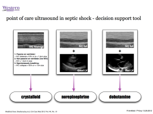
The biggest wrinkle of course is how to manage volume in patients who are triggering the vent and show features of IVC collapse. We know that all the studies to date proving volume responsiveness with mechanically ventilated patients has been done with those passive on the ventilator. It has been our experience, however, that if the IVC itself is flat (<1.5cm in a vented patient) then this is a reliable feature of volume responsiveness. More conservatively, we can say that these patients will be volume tolerant (to use Scott Weingart’s terminology). Further work needs to be done with this and certainly integrating the LV function in these patients as well as their aeration pattern on lung ultrasound (A diffuse A profile would suggest tolerance, as well – as encouraged by Lichtenstein in his FALLS protocol).
Download the pdf of the above support tool here.
]]>See below for a 45 minute panel discussion (hosted by Matt and Mike from Ultrasound Podcast) on the topic of POC ultrasound in the assessment and management of PE. Was conducted and recorded using Google Hangout.
Panel members include luminaries of the field including:
Scott Weingart, Matt Dawson, Mike Mallin, Cliff Reid, Mike Stone
As well, Western Sono’s own Rob Arntfield was invited to join.
]]>
The Critical Care Outreach Team is called to asses a 64 year old male on the thoracic surgery floor complaining of increasing shortness of breath and hypotension. He is status post left upper lobectomy for non small cell lung cancer 1 year ago. He has been re-admitted for workup of a possible recurrence in his mediastinal nodes.
In the 24 hours prior to CCOT arrival he had become progressively acidotic and hypotensive. This was presumed to be on the basis of sepsis and appropriate treatments had been instituted. Despite this the patient remained hypotensive with BP sitting 90’s/50’s. His gases and clinical state worsened and he was taken to the ICU for further evaluation and monitoring.


As is routine for patients admitted to our ICU’s with circulatory failure, a point of care echocardiogram was performed on arrival to the ICU:
Subcostal 4 Chamber
[youtube http://www.youtube.com/watch?v=M9s-KxcycK8?rel=0]
Parasternal Short Axis
[youtube http://www.youtube.com/watch?v=q8YUn6-mR9Q?rel=0]
Apical 4 Chamber
[youtube http://www.youtube.com/watch?v=QWtaWGdMZdA?rel=0]
What is your impression of the images? Is there any significant abnormalities seen? Click HERE for the answers and discussion and to learn the outcome of the case.
]]>[youtube=http://www.youtube.com/watch?v=GSp_xyWBKK0&w=420&h=315]
]]>