Surprising finding in the liver, atelectasis vs pneumonia, and introducing left atrial pressure!
In this episode, we start by discussing the incidental finding of hyperechoic spots in the liver, which we conclude to represent air in the portal system in this patient in septic shock. Then, we move on to the lung with the age old debate of atelectasis vs pneumonia. Finally, we touch on a more advanced application in cardiac ultrasound that is helpful in distinguishing between hydrostatic and non-hydrostatic B-lines; E/e’ to estimate left atrial pressure (LAP).
For questions, comments or feedback, please email us at westernsonopodcast@gmail.com
Post-production by Hubert Gaudreau-Simard
Multimedia support by Brian Park, Meds 2023
Music: Nightchaser by Shane Ivers – https://www.silvermansound.com
Clips
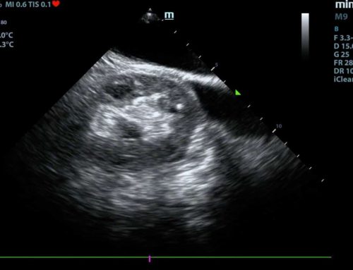
Clip 1: Hyperechoic dots in the liver representing air (apologies for the gain & depth; trainees in action!)
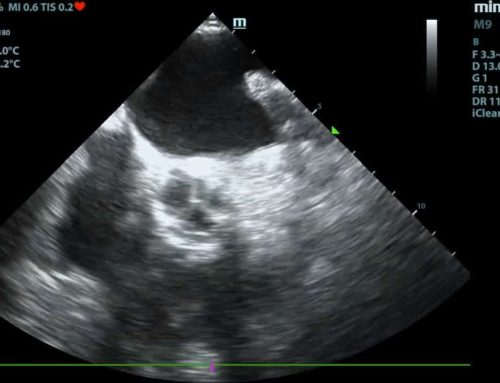
Clip 2: Pleural effusion with consolidation
A) More likely to represent atelectasis
B) Favored to be pneumonia
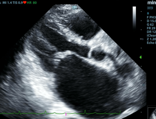
Left Atrial Pressure
Click here to see the detailed PDF!
LAP helps differentiate B-lines caused by hydrostatic pulmonary edema versus inflammatory/infectious causes of B-line.