CCW POCUS crew has been hard at work and would like to welcome this months’ rotators Dr. Kimberley Lewis, Mo Dairi, and Steve Roy. They will be providing some of the COTW cases over the next few weeks (we hope!).
This weeks case comes from Drs. Fulan Cui and Scott Anderson. We’ll leave the interpretation and critique of Andersons POCUS skills to you (but feel free to reply-all… just saying!)
The Case:
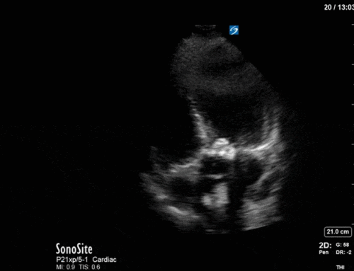
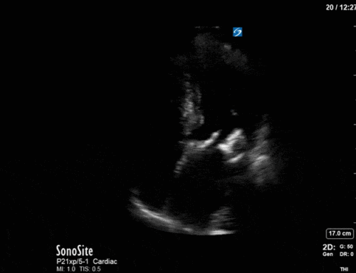
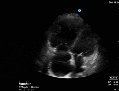
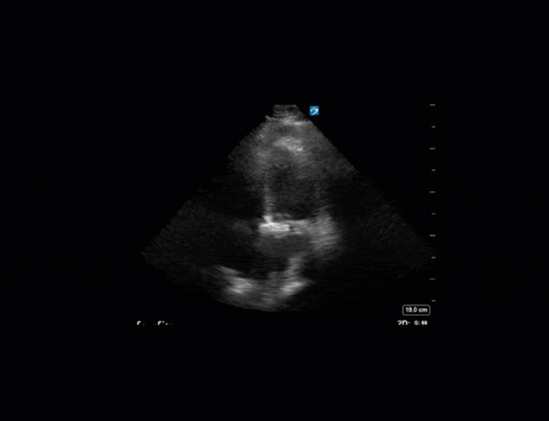
70’s F with known severe aortic stenosis and CAD was admitted to hospital with progressive shortness of breath and was awaiting cardiac surgery. She was brought to CSRU with refractory hypotension, and a POCUS was completed to assist in shock differentiation. Here is one clip: What are we looking at? What is your differential for what this might be?
Last Weeks Case:
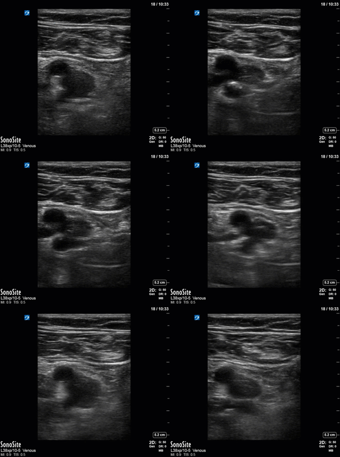
This clip comes from a DVT study. This image was captured using a linear probe on the vascular setting. With the probe marker oriented towards the patients’ right side, we can tell that this is the right leg (NAVY remember? the vein will always be on the same side as the limb). POCUS DVT studies are performed using compressive ultrasonography. In general, depth should be adjusted to see both artery and vein as we see here. Assess the 2D image for apparent clot or thrombus and if nothing is visible apply direct vertical pressure to assess for compressibility. The vein should be compressible when applying less pressure than necessary to compress the adjacent artery. Several anatomic regions need to be assessed, please refer to your favorite POCUS reference for an in-depth review of required anatomy. This clip, taken at the level of the lateral perforator demonstrates clear clot within the vein, and so no compression was applied. This patient went on to be diagnosed with DVT and was placed on therapeutic anticoagulation.
Hope you enjoyed today’s installment of COTW,
The POCUS Team

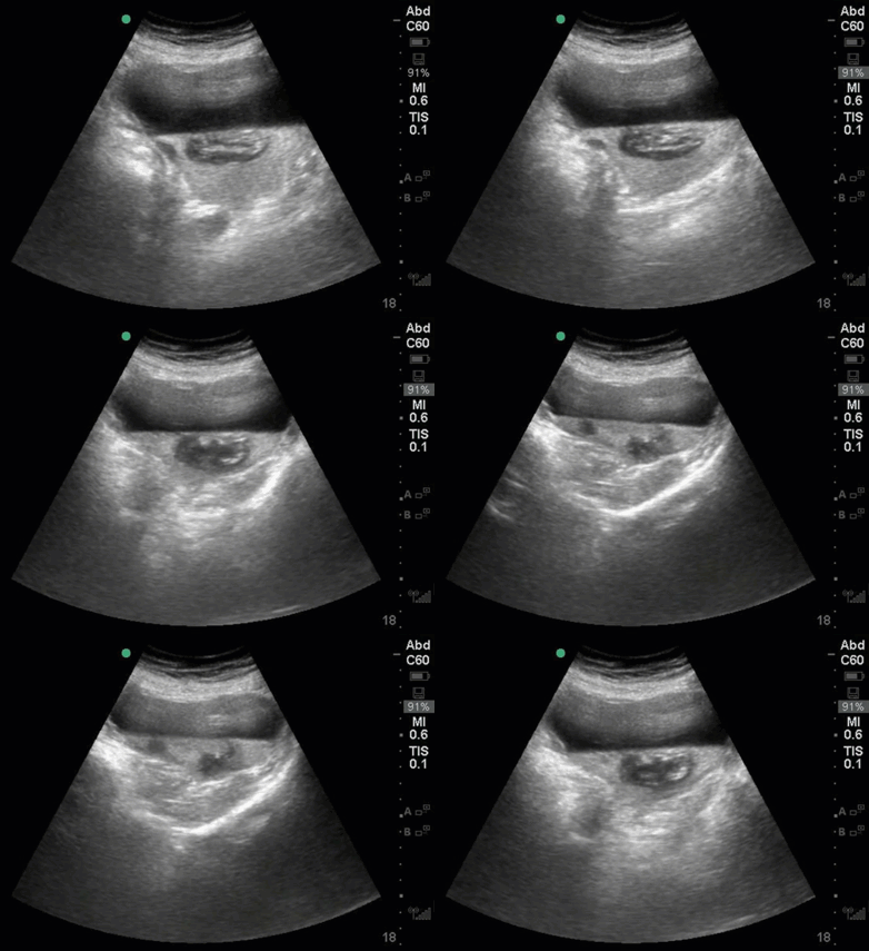
Is that a fluid fluid level and something within the bladder?