Welcome to our newest rotators: Ali AlHarbi, Mike Beyea, and Zainab AlDuhalib who will be providing you with COTW material for Block 12.
The Case:
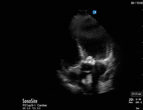
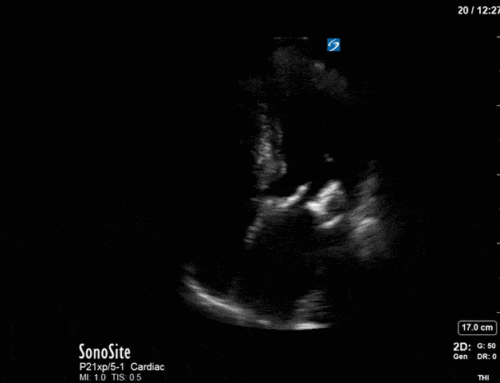
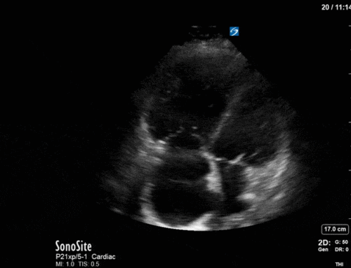
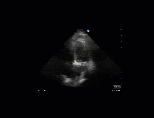
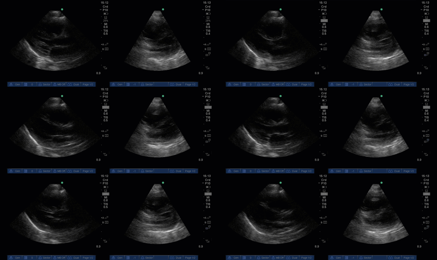
Without any clinical context – can you guess what type of patient this clip might be from? What abnormalities can you spot?
COTG:
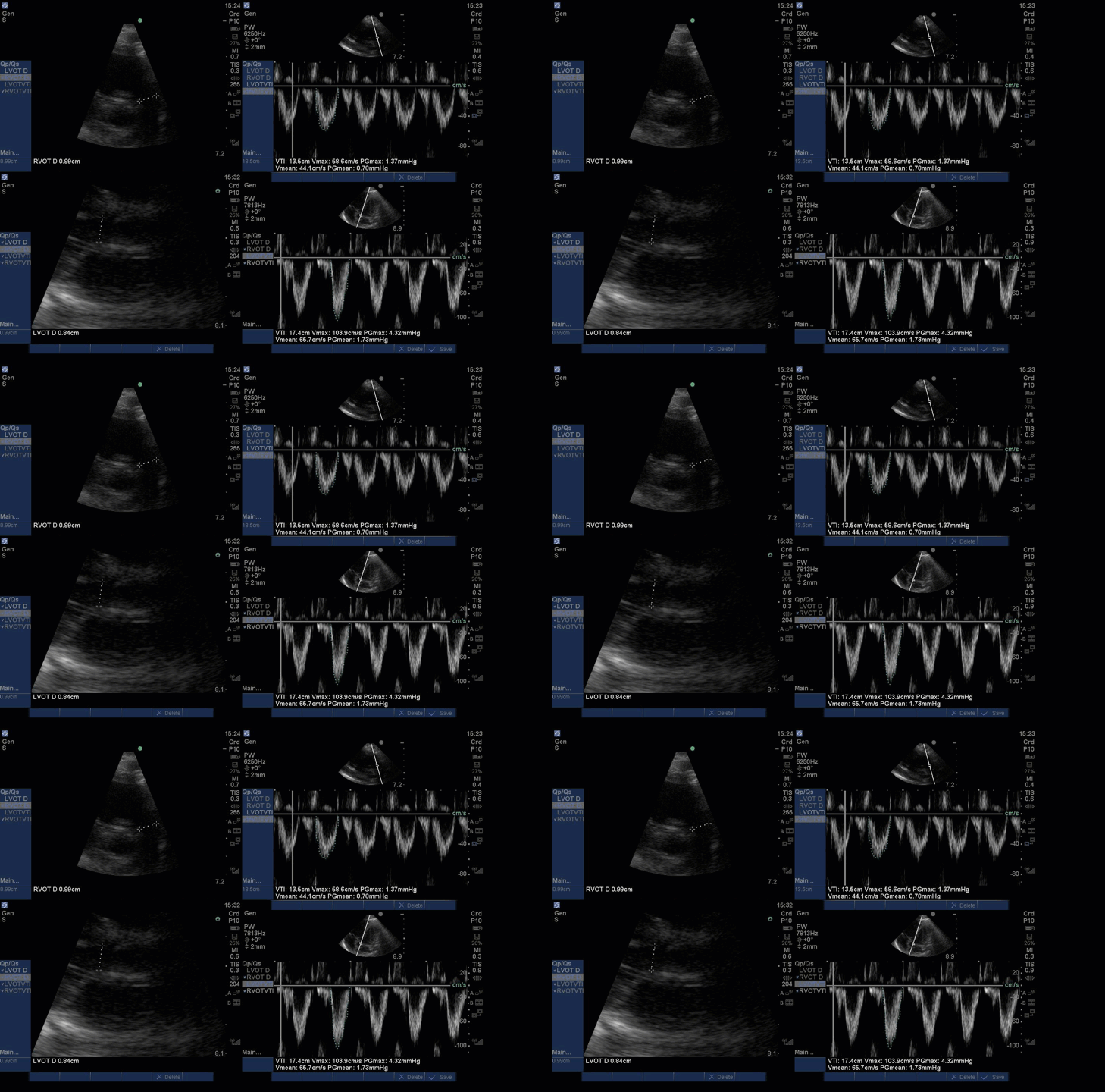
Same patient, what are we assessing for here?
Answer from Last Week:
Last week we presented the case of a 62-year-old gentleman requiring paracentesis. To guide paracentesis a high-frequency linear probe was used to generate the first image of the abdominal wall in the near field with ascitic fluid in the far field. The next still image depicts measurements approximating how thick the abdominal wall is, and how much fluid lies beneath. This offers the proceduralist an estimate of how far the needle should be advanced before fluid will return. Finally, colour Doppler has been used to assess for vessels – most importantly to check for the presence of the inferior epigastric artery which could lead to significant bleeding if lacerated or punctured. The third clip demonstrates pulsatile colour flow, indicating the presence of a vessel and necessitating a different location for paracentesis. Look for the upcoming CHEST article Better with ultrasound – Paracentesis for more info (currently article in press)

The Clip-of-the-Geek:
Also acquired with the linear probe, these clips depict the diaphragm of a patient with a C-spine injury following MVC while he is spontaneously breathing on a trach mask trial. Diaphragm assessment in the critically ill generally involves two measurements: Diaphragm excursion, and thickening. To measure thickening (what we’ve done here), use a high-frequency transducer to locate the diaphragm, which appears as a three-layered structure (diaphragmatic layers of pleural and peritoneal pleura, and a middle hyperechoic layer of muscle). From here use M-mode to take caliper measurements of diaphragmatic thickening at end inspiration, and end expiration. Thickening fraction = (end inspiratory thickness – end expiratory thickness)/end expiratory thickness. Diaphragm assessments in critically ill are very complex and will depend on the patients’ sedation levels, injuries, ventilatory support, etc. In this case, the fact that the diaphragm was moving, and thickening was helpful to rule out diaphragmatic paralysis due to high C-spine injury.

Until next time!
The POCUS Team

