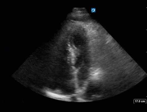
The COTW:
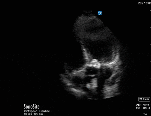
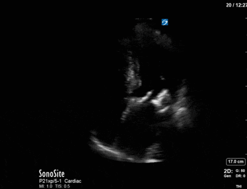
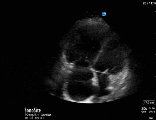
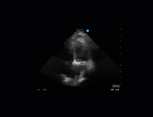
A young male with a collagen-vascular disorder presents with shock. Here is one view from his POCUS, what do you think? Leave a comment below!
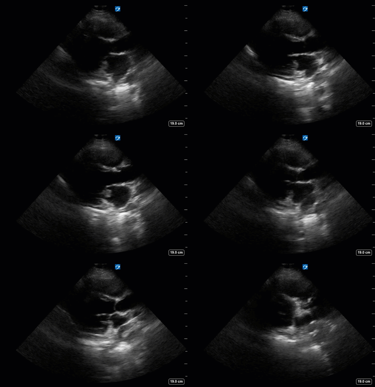
Answer from Last week:
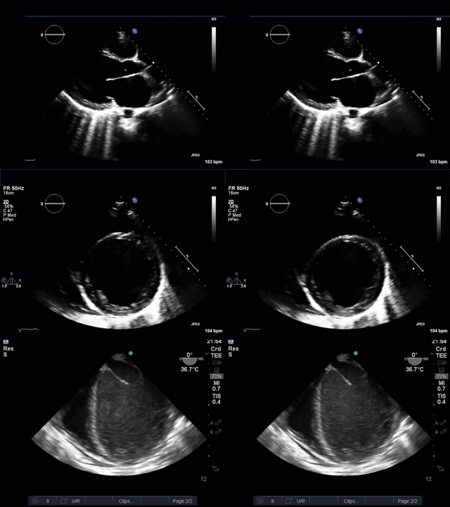
This young man has a very broken looking heart, and it wasn’t because his true love crushed him on Valentine’s day – he has a dilated cardiomyopathy. Vince Lau, our POCUS alumnus and echo-aficionado submitted the case last week as well as these teaching points:
1) LAE size (severely dilated)
– When Brian Buchanan (former POCUS fellow) was here, he liked to state the phrase that the triscupid valve for regurgitation was like a “canary in the coal mine” (acted like an early warning system)
– If the TV is a “canary in the coal mine”, then the LA enlargement is like the “boy who cried wolf, but the sheep are already eaten” (more like a late warning system – sorry for the morbidity/mortality of this statement)
– Explanation: LAE is a sign of chronicity of a disease process (i.e. long-standing HTN, AS, chronic MR/MS, etc.)
– So in essence, if LAE is seen, it means that it’s been going on a while (is a late finding), and there maybe other findings of chronically elevated LAP (i.e. pulmonary HTN, RV dilation, TR)
– In contrast, and acute process that causes elevated LAP (i.e. acute MR caused by chordal rupture) will likely not have LAE acutely
2) Spontaneous echo contrast (“smoke”) – nidus for clot
– “Smoke” primarily in the LV during POCUS TEE – in comparison to other chambers
– LV thrombus found intra-op during LVAD insertion, not seen on prior TTE images by cardiology (no Definity given)
– Note the lack of decompression/off-loading of the LV (still massively dilated) despite LVAD insertion (likely secondary to location/position of in-flow cannuale) – offloading LA instead of LV directly

