
The Case
This was a case of an 80 yo M, previously healthy, with no known significant previous medical history. He presented to the ED with acute decreased level of consciousness. The leading concern was acute spontaneous ICH. He was intubated and taken for a CT Head which surprisingly was completely normal. He was subsequently transferred to the ICU with the diagnosis of altered LOC NYD. A POCUS study was performed, and several cardiac clips are shown below. What do you think? Does this give a potential clue to what could be going on? 


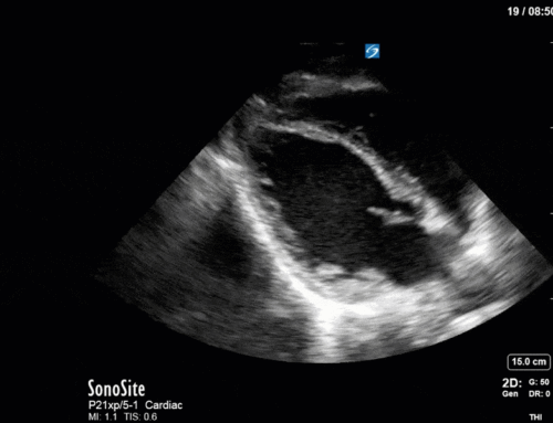
These clips show significant multivalvular pathology. The most obvious striking abnormality is the significant hyperechoic structure mostly localized on the posterior mitral valve leaflet (PMVL). Although this has a differential, this was favoured to be severe mitral annular calcification (MAC). Given the size and significance of the MAC there was further concern that this may be have provided substrate for an embolic basilar artery stroke as the cause of his decreased LOC. He was taken back for an immediate CTA which again was normal. However, the next day, an MRI was performed which unfortunately showed catastrophic multifocal, bilateral infarcts involving cortical, subcortical and brainstem structures consistent with an extensive cardioembolic etiology. Although this was an unfortunate outcome, this case still captures the power of POCUS to shed light on what was very much an undifferentiated patient with altered mental status. The study prompted further consideration of a cardioembolic stroke and expeditated the necessary investigations. Furthermore, hemodynamically significant valvular lesions (see below) were also recognized which allowed the team the alter their management to keep the patient stable while further investigations were completed.
As a result of the PMVL pathology there is at least moderate mitral regurgitation which is only partially captured in the above clips. Furthermore, you can also notice flow acceleration through the MV suggestive of mitral stenosis. Interrogation with CW (PW was aliased in this case) shows the very high (~2m/s) mitral inflow E-wave velocities. We can then trace the mitral inflow to get a mean gradient which in this case was approximately 7 indicating moderate mitral stenosis.
The aortic valve is also heavily calcified with very limited opening highly suggestive of significant stenosis. Thus, this was also interrogated with Doppler to further quantify its severity. First, PW Doppler was used to measure the LVOT VTI which was around 18cm. Next, CW was placed through the AV, which when traced, gave an AV VTI of 87cm, a peak velocity of 4.2m/s, and a mean gradient of 34mmHg. The dimensionless index (LVOT VTI/AV VTI) was therefore 0.21. Although the mean gradient of 34mmHg is in the moderate category the rest of the parameters converge on this being severe AS. Thus, POCUS was able to quickly discover severe multivalvular pathology that was not previously documented.
This case again highlights how the deployment of POCUS can uncover significant pathology which can dramatically alter the hemodynamic management of patients, and simultaneously aid in determining the ultimate diagnosis of the patient.
Thanks for reading!
Matt


Thanks for sharing this case, very good explanation .
This is not only a great example of sweet sweet pocus goodness, but of the limited value of non contrast head CT in acute decreased level of consciousness. ICH often give you enough time to have some clinical signs (headache, seizure, lateralizing signs) before dropping their LOC. Brainstorm infarcts, not so much. If you’re taking these guys to ct, stroke protocol them!