
Hi POCUS people,
This week we have a special treat for you as we head downstairs to the depths of the sleepless and always on-the-go Emergency Department, with two great cases from our WesternSono Emergency Medicine contingent. The COTW was written by Dr. Aaron Stone (Western Sono ED POCUS fellow) and the COTG was written by Dr. Melissa Snyder (Western Sono ED POCUS fellow), based on a case by Dr. Frank Myslik (Western Sono Emergency Medicine Ultrasound Fellowship Director).
The Case
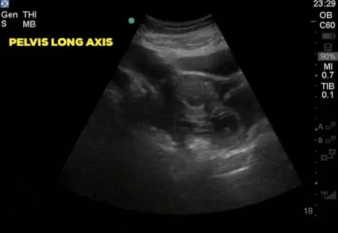
A 30-year-old female, presents to the emergency department with acute lower abdominal pain. She is 9 weeks pregnant by dates and has had no formal ultrasound prior to presentation. Her initial vitals are; HR 102, BP 116/70, RR 24, T 36.2 and SpO2 98% on RA. A bedside ultrasound is completed and demonstrates:
COTG
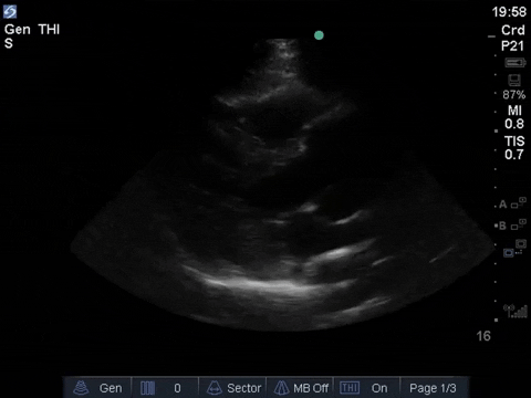
A 57 year old male presents with sudden-onset headache and pain in his chest, lower abdomen and back. He also notes a few episodes of diarrhea that morning. He is Vitals: 121/75, HR 69bpm, 36.5deg C, RR 21, 99% on room air. You put the probe on the patient’s chest and this is what you see:
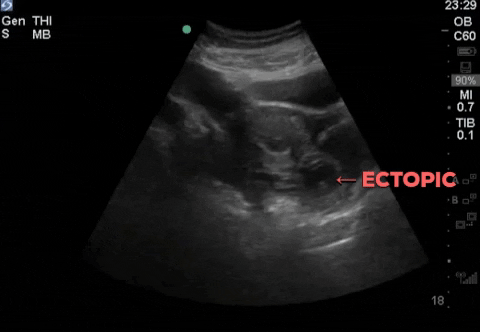
POCUS of the patient’s RUQ is shown here:
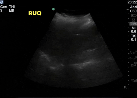
Initially the most likely diagnosis is an ectopic pregnancy. A live fetus can been seen posterior to the uterus in this longitudinal view of the pelvis. Following the identification of an ectopic pregnancy the RUQ and LUQ views can be obtained to assess for free fluid. The second clip demonstrates free fluid in the hepato-renal interface. This has now raised our suspicion from an ectopic pregnancy to a ruptured ectopic pregnancy. The ultimate disposition is the OR. Obs/Gyn should be consulted immediately and prior to obtaining the results of further imaging. The patient should be placed in an acute care setting and 2 large bore IV access should be obtained.
The presence of free abdominal fluid by POCUS has been shown to predict the need for operative intervention, when an ectopic pregnancy is suspected. Moore et al. completed a prospective study on 242 patients suspected of having an ectopic pregnancy and found:
- Free fluid in the hepato-renal interface had a sensitivity of 50%, specificity of 99.5%, positive LR of 112 and a negative LR of 0.5 for the need of surgical intervention in suspected ectopic pregnancy.
- Free fluid in the pelvis had a sensitivity of 56%, specificity of 94%, positive LR of 9.5 and a negative LR of 0.47 for the need of surgical intervention in suspected ectopic pregnancy.
- Adding the RUQ and LUQ views to a confirmed ectopic or NDIUP POCUS scan can be a game changer and is strongly encouraged.
Further reading:
Moore C, Todd WM, O’brien E, Lin H. Free fluid in Morison’s pouch on bedside ultrasound predicts need for operative intervention in suspected ectopic pregnancy. Acad Emerg Med. 2007;14(8):755-8.
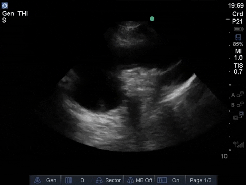
This single parasternal long axis clip should make you very concerned for an aortic dissection. The first and most glaring thing you might notice is the violation of the “rule of thirds”. In the PLAX view, typically the RVOT, aortic root, and the LA are all about equal in diameter. In this clip the aortic root appears dilated. The second clue is a mobile, linear, and hyperechoic structure that appears in the descending thoracic aorta (in the far field). It appears consistent with a dissection flap but must be confirmed on additional views to ensure it’s not an artifact. Although the presence of a flap is consistent with an aortic dissection, importantly with POCUS its absence does not rule out the possibility of aortic dissection. In this case it would also be useful to assess for aortic valve regurgitation and presence of pericardial effusion, both complications of Type A aortic dissections.
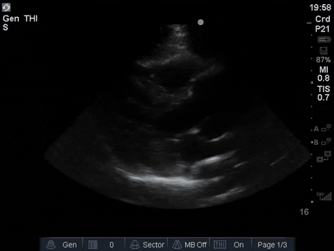
The suprasternal view is a useful view when looking for Type A aortic dissections as it shows the ascending aorta and it’s arch. Typically it’s first three branches (brachiocephalic artery, left common carotid artery and left subclavian artery) are seen in the near field of the screen. Watch this video by Jacob Avila of 5minsono.com and The Ultrasound Podcast for a quick review of POCUS for aortic dissection.
In 2017 Gibbons et al, described a protocol for detecting aortic dissection. The presence of one of: pericardial effusion, aortic intimal flap, or outflow tract diameter at end-diastole >3.5cm showed a sensitivity of 100% for Type A aortic dissections and 96.4% for all dissections. POCUS can be helpful in expediting the diagnosis of aortic dissection as was shown in this case. Pare et al (2016), found that POCUS reduced time to diagnosis by over 2 hours. Delays in diagnosis of aortic dissection are associated with increased mortality rates.
Further reading:
Gibbons R, Smith D, Mulflur M, et al. Bedside point of care ultrasound for the detection of aortic dissections in the emergency department [abstract] Ann Emerg Med. 2017;70(4):S143.
Pare JR, Liu R, Moore CL, et al. Emergency physician focused cardiac ultrasound improves diagnosis of ascending aortic dissection. Am J Emerg Med. 2016;34:486–92.
Happy scanning,
Marko Balan, Aaron Stone, Melissa Snyder, Frank Myslik on behalf of the Western Sono team


