
Hi POCUS enthusiasts,
We had some great participation with our inaugural interactive COTW/COTG, so keep posting! Thanks for everyone who signed up for the our web-based COTW/COTG personalized emails. For those who have yet to sign up just go to westernsono.ca and insert your name and email into the text box on the right side of the webpage. Then get ready to flex your POCUS muscles and post your answers/votes.
Or click here to access the case immediately:
The Case
A 42 year old female is admitted with ARDS and a pleural POCUS is performed. Due to difficulties identifying lung sliding, M-mode was used to evaluate for pneumothorax. Based on this image, does the patient has a pneumothorax? What is the 2D ultrasound image correlate of the vertical areas as shown by the arrows?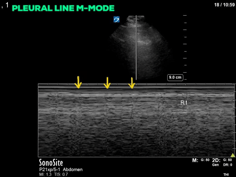
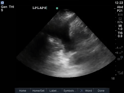
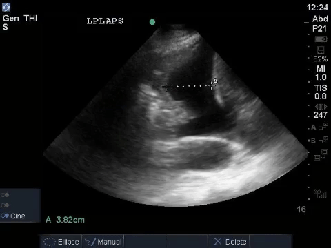
In the same patient the following images were obtained on the contralateral side.
COTG
Answer to Last Weeks Case
A middle aged man presented to the Emergency Department 5 days after a ERCP and stent insertion for obstructive jaundice feeling unwell, feverish, and lightheaded. He rapidly deteriorated with hypotension and was admitted to the ICU with sepsis NYD. What (if anything) is concerning on this patient’s RUQ/biliary POCUS? What management steps would you recommend based on these images?
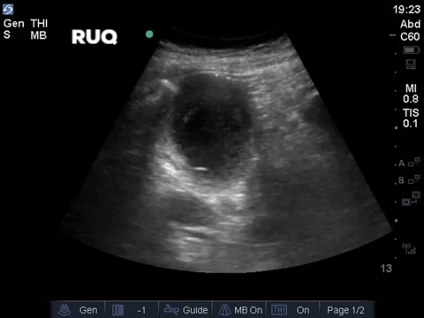
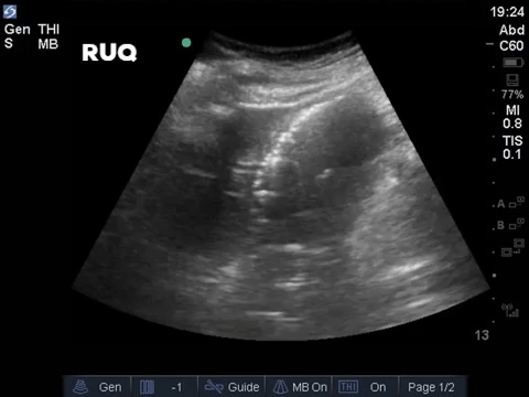
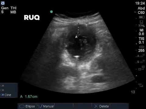
Great work by all the respondents online! You were right on with your interpretations. These images show small hyperechoic “dots” throughout the gallbladder. Although you might initially think of stones or polyps when seeing hyperechoic structures within the gallbladder, these have the typical appearance of small locules of air (almost like an air bronchogram). The trick here is to recognize that these don’t create acoustic shadowing, as a stone(s) would, and they are small and numerous which would not be expected for a polyp. In the last image the gallbladder wall measures nearly 1.6cm!!! This is way beyond the normal thickness of up to 3mm. Other POCUS findings in acute cholecystitis include a sonographic Murphy’s sign and pericholecystic fluid.
Based on these POCUS findings the patient had an urgent cholecystotomy tube inserted for source control of his septic shock. Cultures of the fluid eventually were positive for polymicrobial growth with multiple gram negative coliforms and enteroccoci species.
Answer to Last Weeks COTG
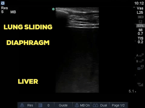
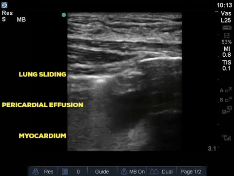
A 32 year old male cyclist vs vehicle trauma patient is hypotensive in the trauma bay. The junior resident shows you these clips and is gearing up for bilateral chest tube insertion. Where do you tell the resident to place the tubes? Also, on the left chest, what is the anechoic space that the arrow is pointing to?
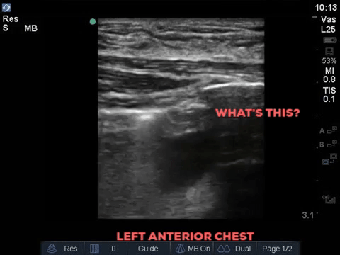
Apologies to everyone but for this one but given your POCUS prowess, we had to pull some tricks out of our sleeve for this one. Neither of these actually represent a lung point, they are both examples of “pseudo-lung point”. On the right side, is the confluence of the lung-diaphragm/liver. Look closely here, the giveaway is that you can see the actual thickness of the diaphragm and the keen eye will catch some more echogenicity in the far field (that’s the liver). On the left side we have another “pseudo-lung point” this time made up by the lung-pericardial interface (this patient actually had a pericardial effusion). To avoid falling into this trap on your next trauma patient, be aware of where your probe is placed when identifying the lung point and have a look with more depth to make sure you don’t miss any important clues (like a liver). Kudos to the 3 voters on the website who didn’t fall for this common and potentially dangerous pitfall! Other potential false positive lung points can be observed in patients with pleural calcification, asbestos-related pleural disease, bullous disease.
Right Side
Left Side
Attached is a brief case report of another pseudo lung point finding in a post-CABG patient. Steenvoorden2018_Article_LungPointInTheAbsenceOfPneumot
Take Home Points
- POCUS findings of acute cholecystitis include: gallbladder wall thickening (>3mm), pericholecystic fluid, and sonographic Murphy’s sign.
- Emphysematous/air artifact creates small hyperechoic points and be weary when this is seen in areas where air should not exist.
- Beware of the pseudo lung point which can be caused by lung-diaphragm interface, bullous disease, and other pleural-based diseases.
Thanks,
WesternSono POCUS team


