Hi everyone,
Happy 2020! Hope everyone had a great holiday! It’s great to be back at it with more great cases!
The Case
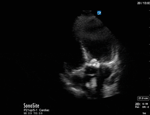
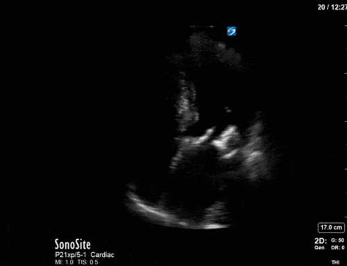
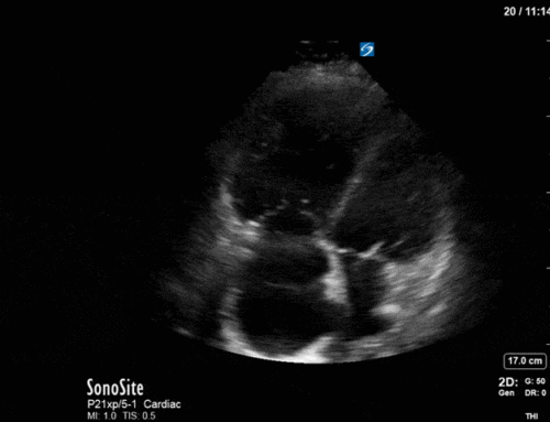
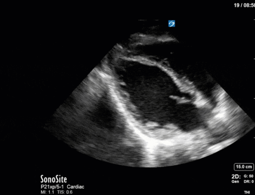
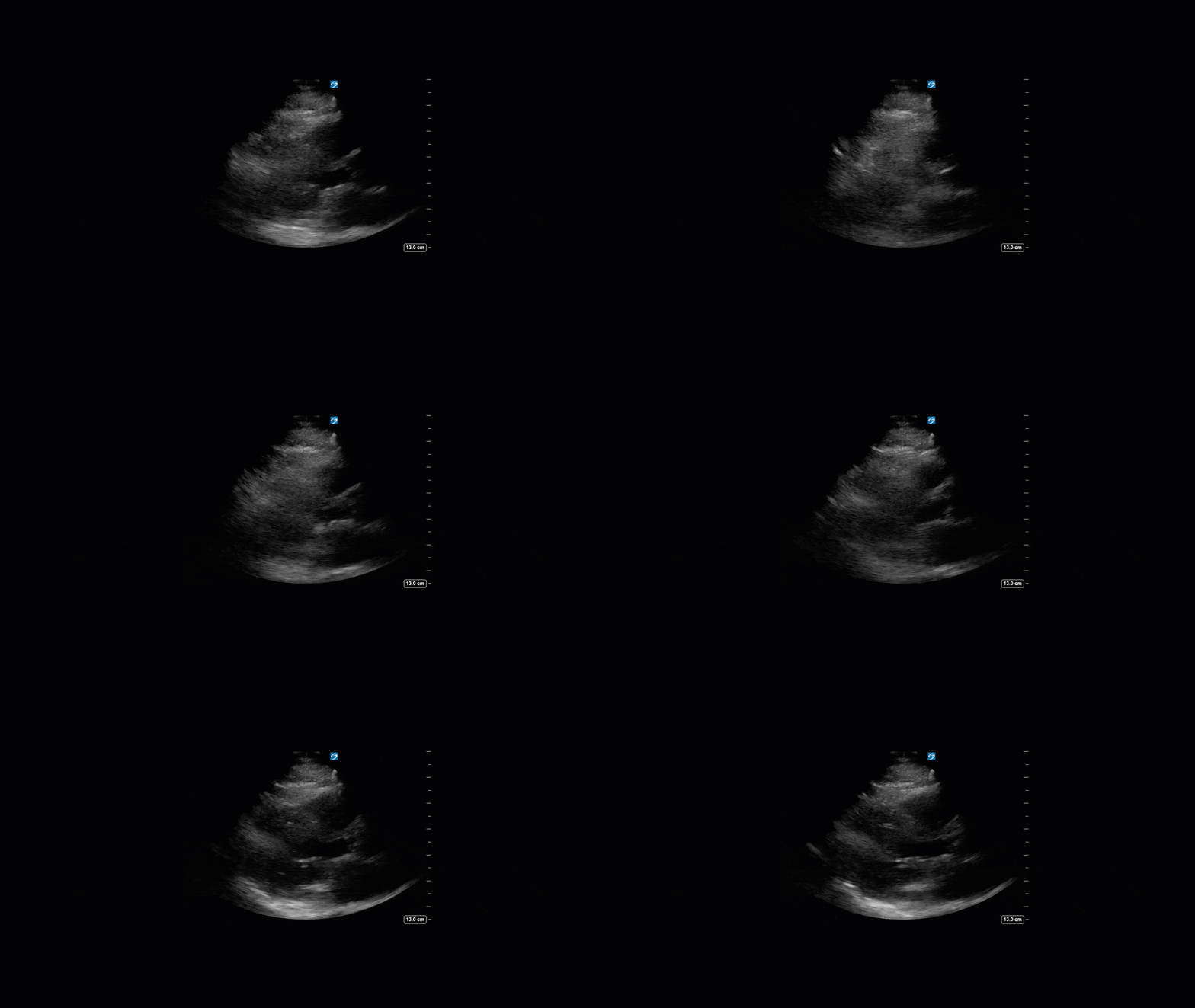
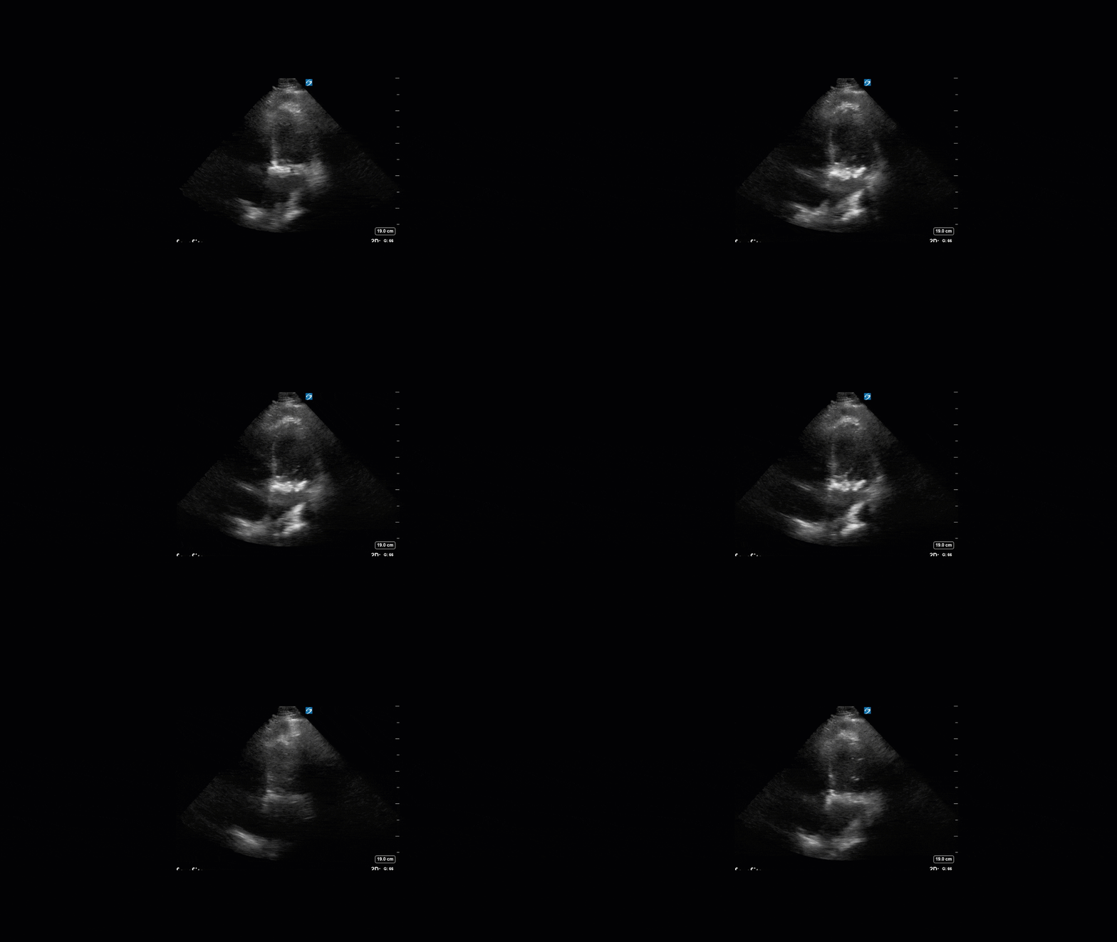
This is a 74yo F with a past medical history significant for ESRD and severe PVD who was transferred to the ICU overnight with presumed septic shock thought to be related to left foot osteomyelitis. She had presented to the ED with refractory hypotension and altered LOC necessitating intubation and high dose vasopressors. The POCUS team went to do a focused cardiac exam, primarily to see if there was a cardiogenic component to his shock. The following TTE images were taken and the decision was made to perform a point-of-care TEE. What do you think is going on?
TTE Views
TEE Views
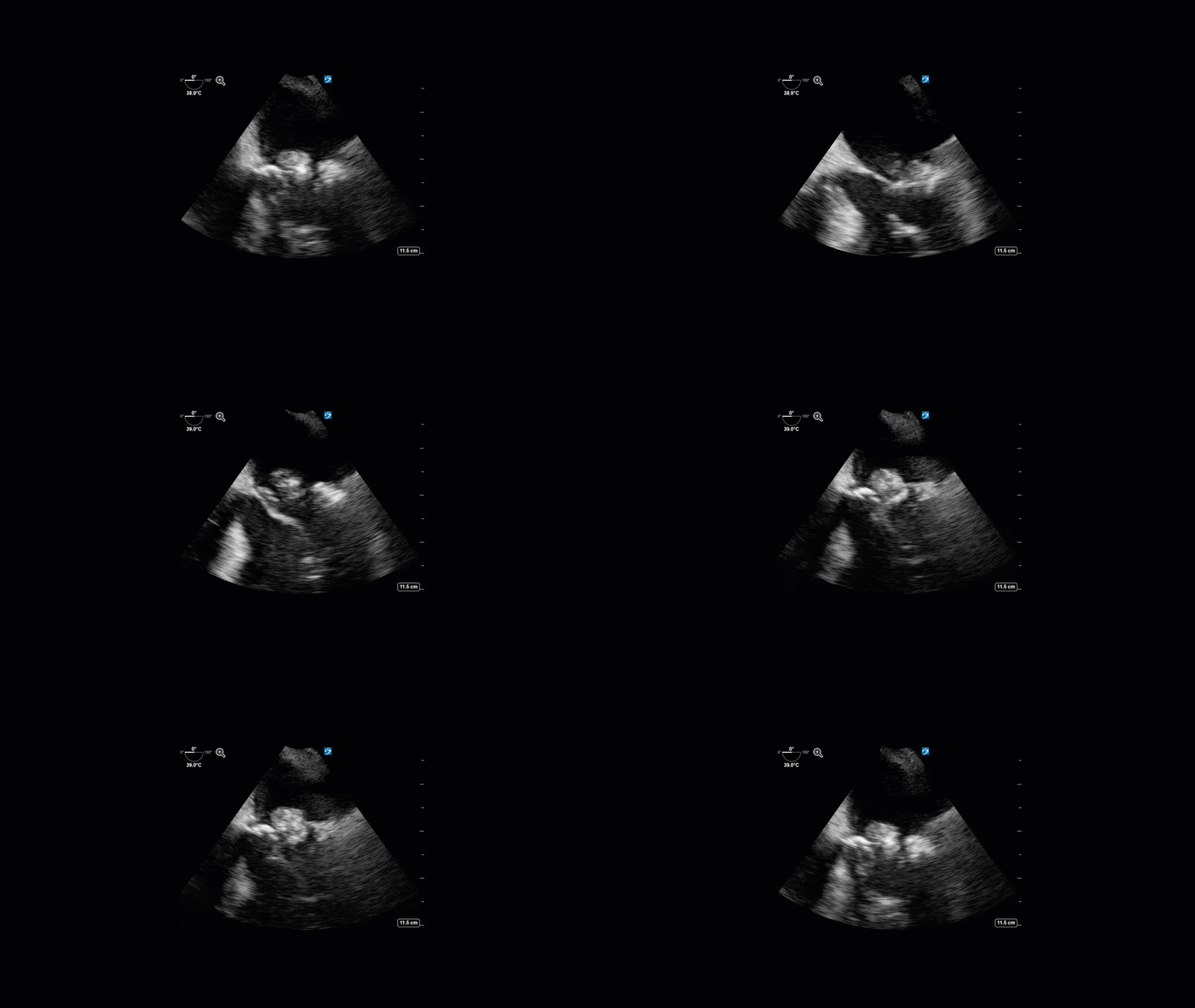
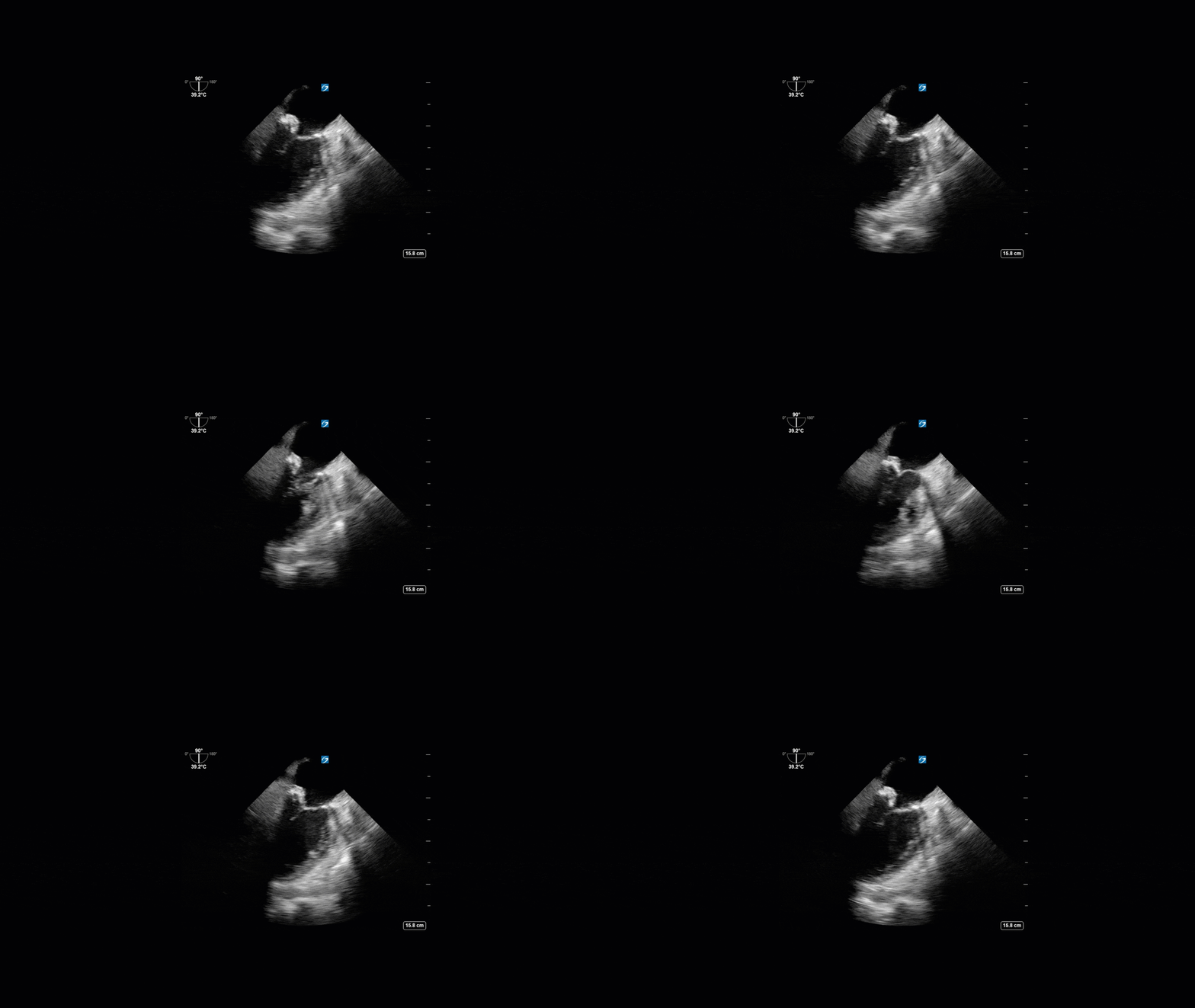
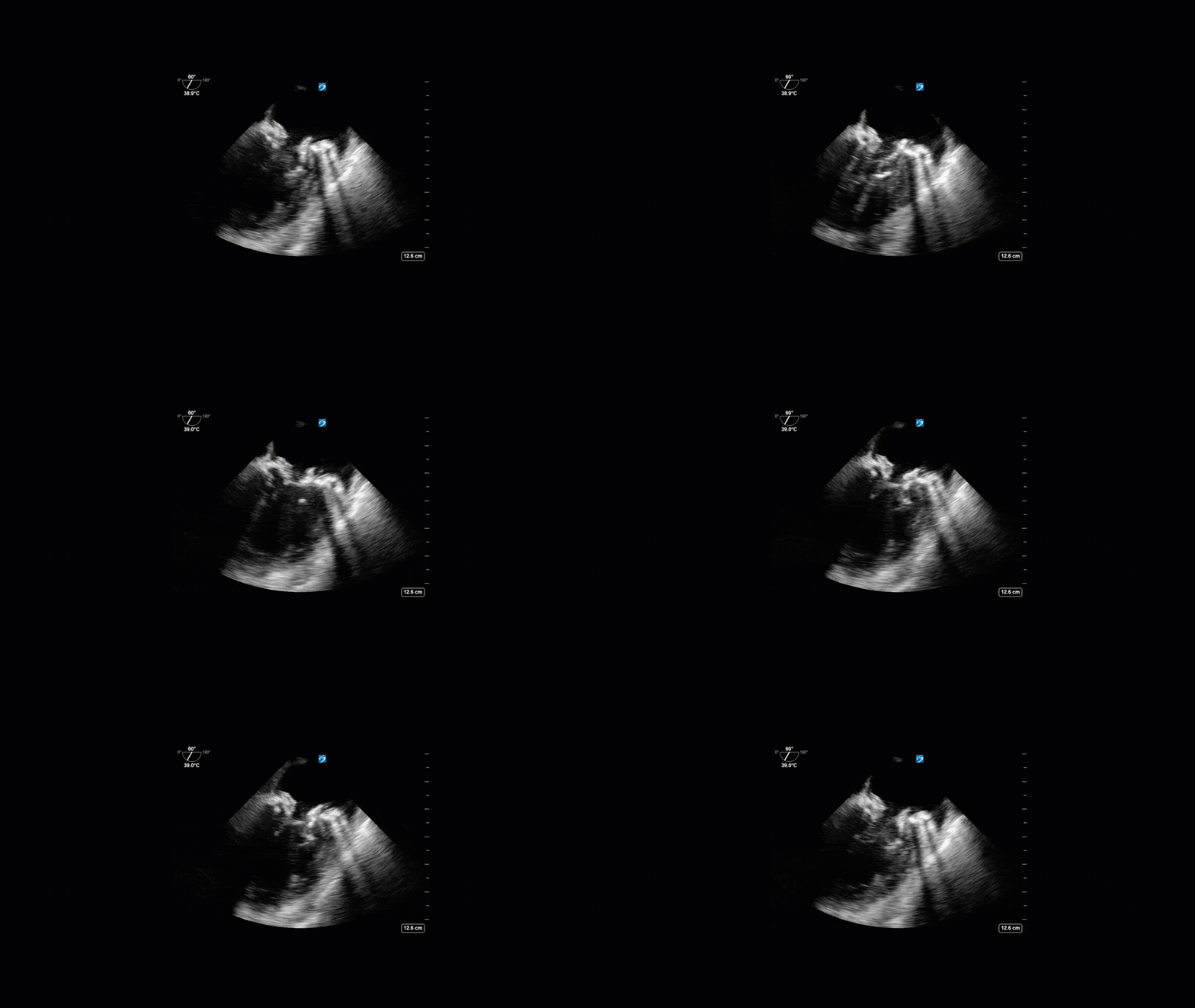
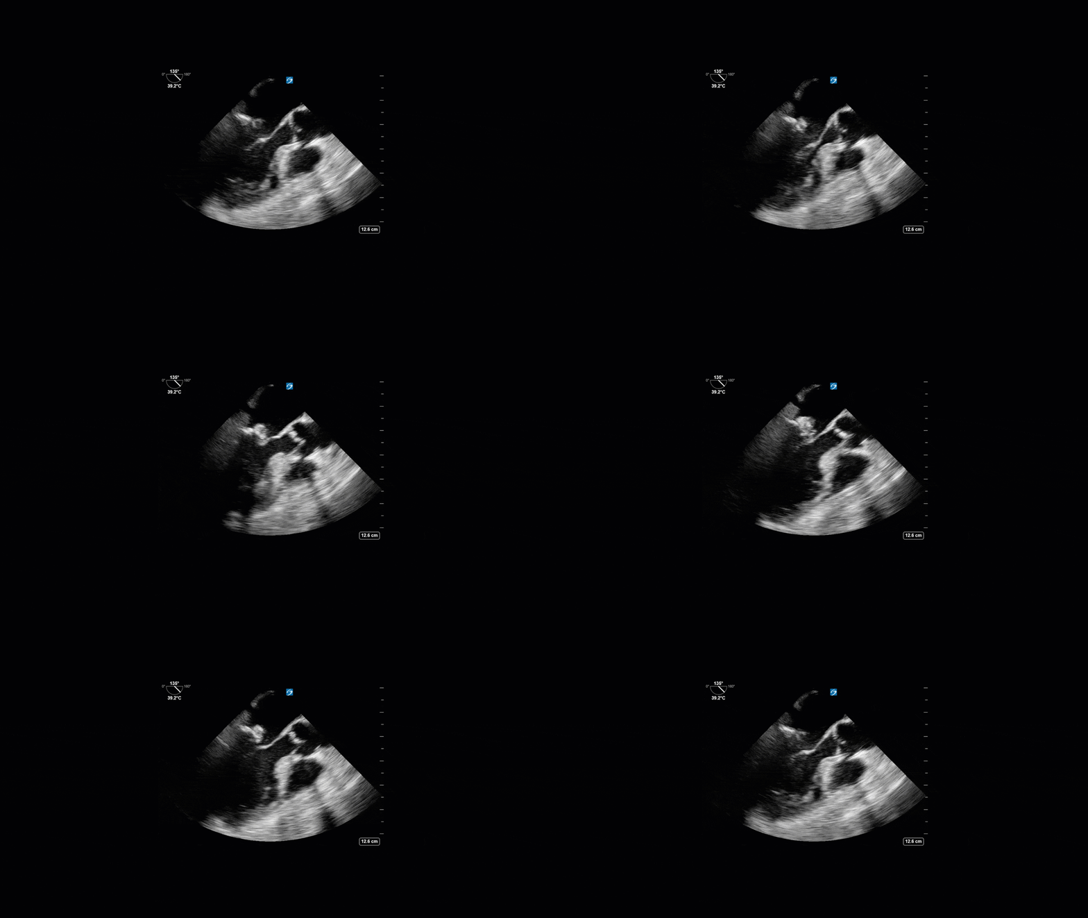
The TTE images clearly demonstrate a grossly abnormal mitral valve, but it’s hard to delineate exactly what’s going on. Dialysis patients (as this patient was) are often predisposed to significant mitral valve annular calcification (MAC). When initially assessing this valve, it was difficult to confidently distinguish between severe MAC and potential endocarditis (although highly suspicious for this).
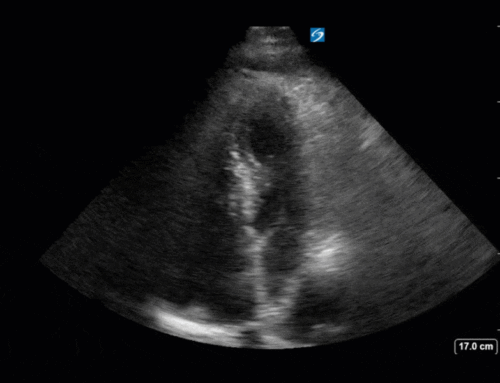
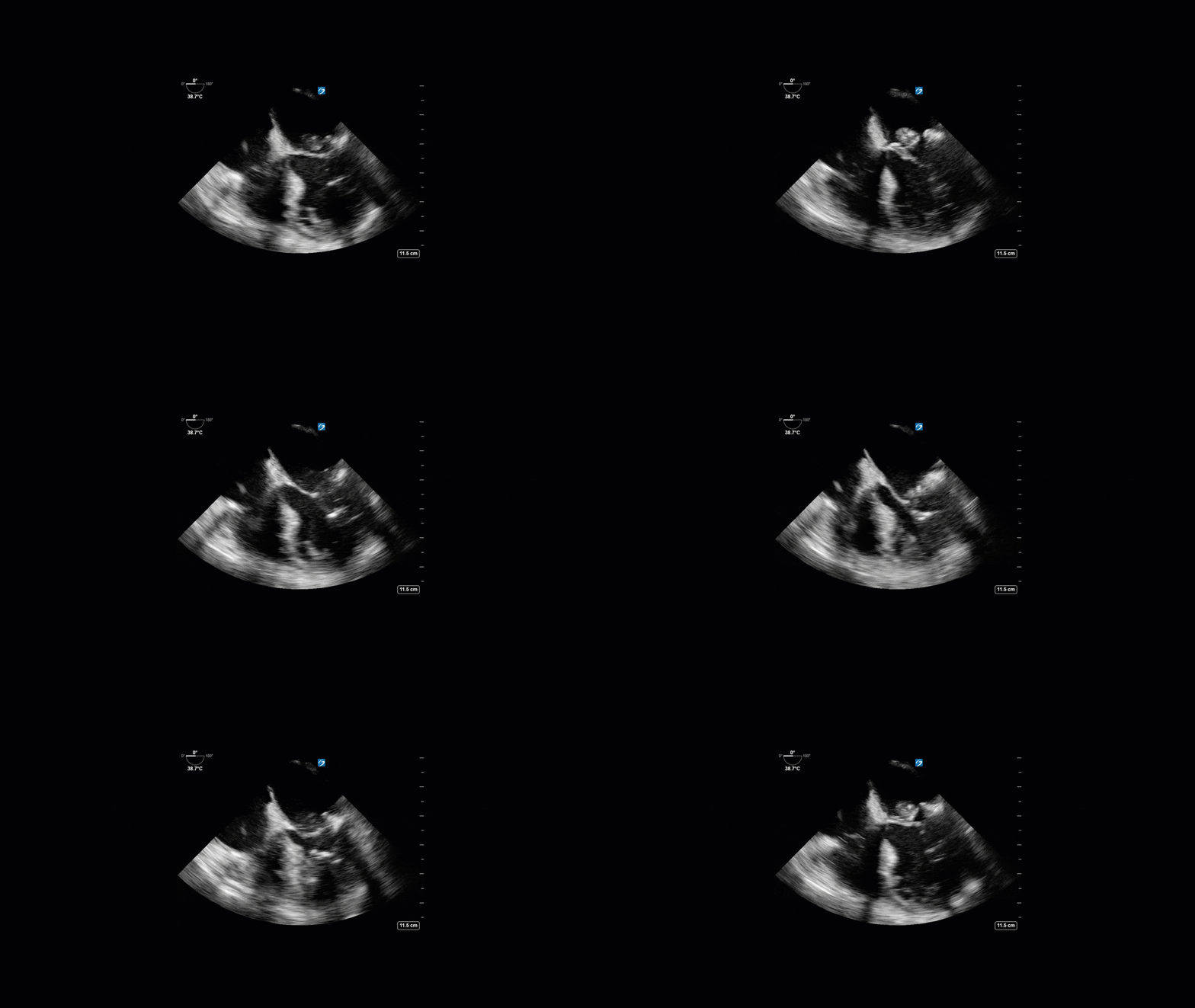
Luckily, we have ready access to a TEE probe, so we quickly made the decision to have a closer look at the valve with TEE. As you can see from those images, the MV pathology is much better delineated and we can assess the MV (and its various scallops) using different omniplane angles. Now we can see a large, independently mobile, hyperechoic lesion primarily on the posterior mitral valve leaflet (although there also looks like there may be some smaller lesions on the AMVL as well). Finally, you might also have noticed a few lesions on the NCC of the aortic valve. Given the pathology evident on the MV, these would also be highly suspicious for AV endocarditis as well (although could also represent calcification).
In the end this patient would grow MRSA in her blood which ultimately would have prompted the team to order an echocardiogram to look for endocarditis. But, using POCUS TEE the patient’s endocarditis was diagnosed more promptly which gave the treating team more diagnostic confidence, avoided chasing other potential diagnoses, and facilitated expedited consultation and management with the appropriate services.

