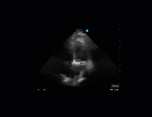
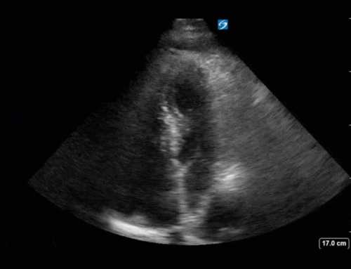
The Case


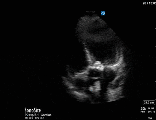
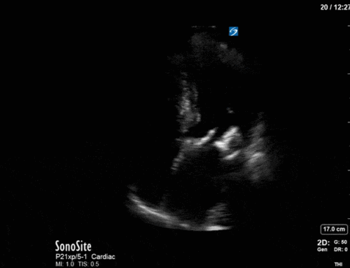
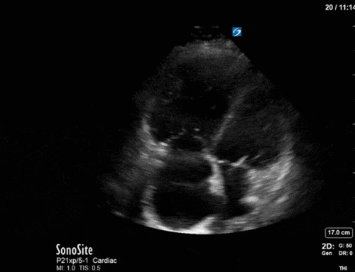
I’m sure you all saw the strange anechoic structure above the diaphragm on the L3 and L4 views. Given the clinical history you might be tempted to think there might be something strange going on (e.g. could it be a lung abscess?). However, when you see things like this, that look a bit strange, you should try to focus your ultrasound field on it and capture it as best you can. We did this as shown below.

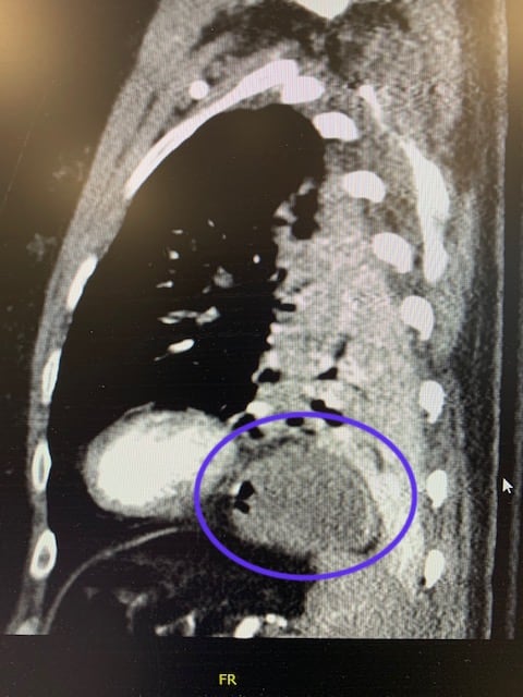
When you focus on it this way you can clearly see the distinct rugae of the stomach! You can even potentially see the NG tube in the center. Thus, this patient just had a hiatal hernia. We saw she had a CT Chest a week earlier (on admission, to ensure that the etiology of her arrest wasn’t a PE), so we quickly looked at that to confirm (image below). If you were still uncertain you could even try flushing the NG tube while looking with US. Moral of the story is sometimes with POCUS we will see things that look unusual, and it’s important that we try to figure out what’s going on, and avoid overcalling or labelling things when we’re not sure. Thanks for reading and stay tuned for next week!
