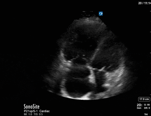
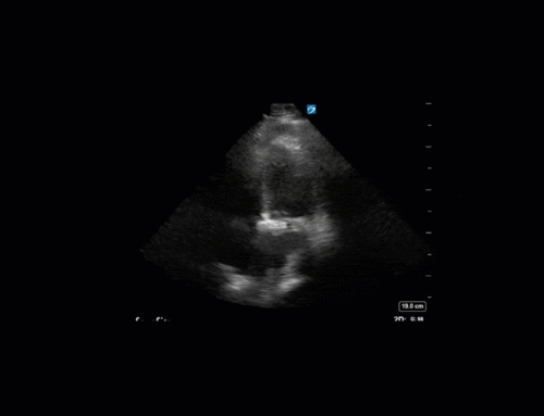
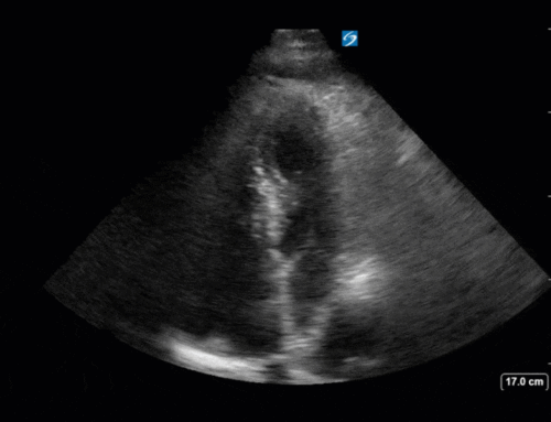
The Clip-of-the-Week Returns this week after a short hiatus in May. To those of you who were worried, we’re sorry, and you can breathe a sigh of relief now because the POCUS cases will continue!
The Case:
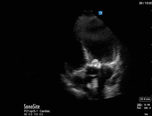
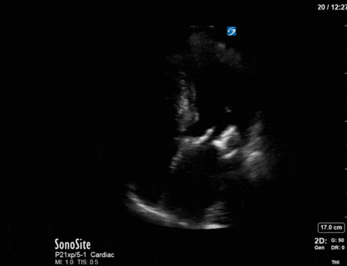
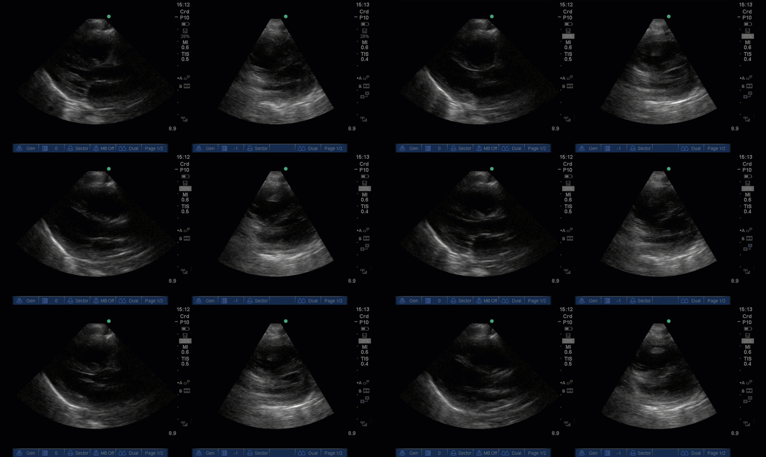
This case comes from Zainab Al Duhalib, thanks Zainab for capturing the clip. Too much context would give it away, but I will tell you that the lung ultrasound showed A-line pattern. What part of the body is this clip from? Which probe was used to acquire it? What do you see?
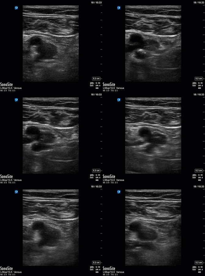
The Last Case:
If you hadn’t guessed based on the depth (only 9cm to see the whole heart and then some!) This was a pediatric case, with the images captured by one of our pediatric critical care colleagues Ali AlHarbi. These images are from a 2-year-old with autoimmune condition NYD. The patient was intubated and ventilated with HFO ventilation. As you can see here the RV appears dilated, with D-shaped septum.
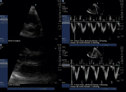
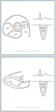
The clip of the week/geek is not the proper venue for a deep dive into pediatric congential echocardiography, but one thing we can try to assess for is shunt which was on the differential in this case. To assess for shunt with echo, we can use a bubble study, or colour Doppler, but a more quantitative/sophisticated measure is a Qp/Qs or shunt fraction. Normally, the right and left heart should have precisely the same stroke volume. When a shunt causes blood to move freely from one side of the heart to another there will be a stroke volume discrepancy. Qp/Qs takes the ratio of stroke volume from the right (pulmonic circulation – p) and the left (systemic circulation-s) to determine the presence and direction of a shunt. Qp/Qs >1 indicating left to right shunt, and <1 indicating right to left. To gather this data, we use the pulmonary artery (obtained in a basal short axis view) to calculate a RVOT VTI in a similar manner to the way we measure LVOT VTI using pulsed-wave Doppler. In this case the ratio of stroke volume was 10.38mL/9.63mL = 1.08, and with this value it is unlikely that there was any significant shunt.
