
Hi Everyone! First, let me introduce myself. My name is Matt White and I am one of the new Critical Care Ultrasound Fellows at Western. I completed my EM and ICU Fellowship at Queen’s and am incredibly excited to be joining the WesternSono team for the year. I am also excited to reignite the COTW. Through this outlet I hope to highlight interesting cases that demonstrate the many ways POCUS is being used to improve patient care in the ICU. There will be a mixture of content from the more bread-and-butter applications to getting into the nitty gritty of advanced spectral doppler. Through this variety, we hope to demonstrate the breadth of a whole-body ultrasound approach to critically-ill patients. With that out of the way, let’s get things started!
The Case
This is a 35 yo M PWID who presented with a right septic AC joint, MRSA bacteremia and hypoxic respiratory failure. He was taken to the OR for washout of his AC joint and subsequently transferred to the ICU for post-op management. A post-operative CXR showed some patchy consolidations but no obvious pleural effusions. The POCUS team was subsequently deployed. Interrogation at the costophrenic angle and PLAPS (posteroalveolar and/or pleural syndrome) point on both sides yielded the following images. What do you see and what should the next steps in management be?
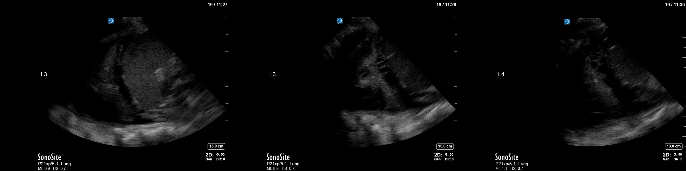
Left Sided Clips
Right Sided Clips
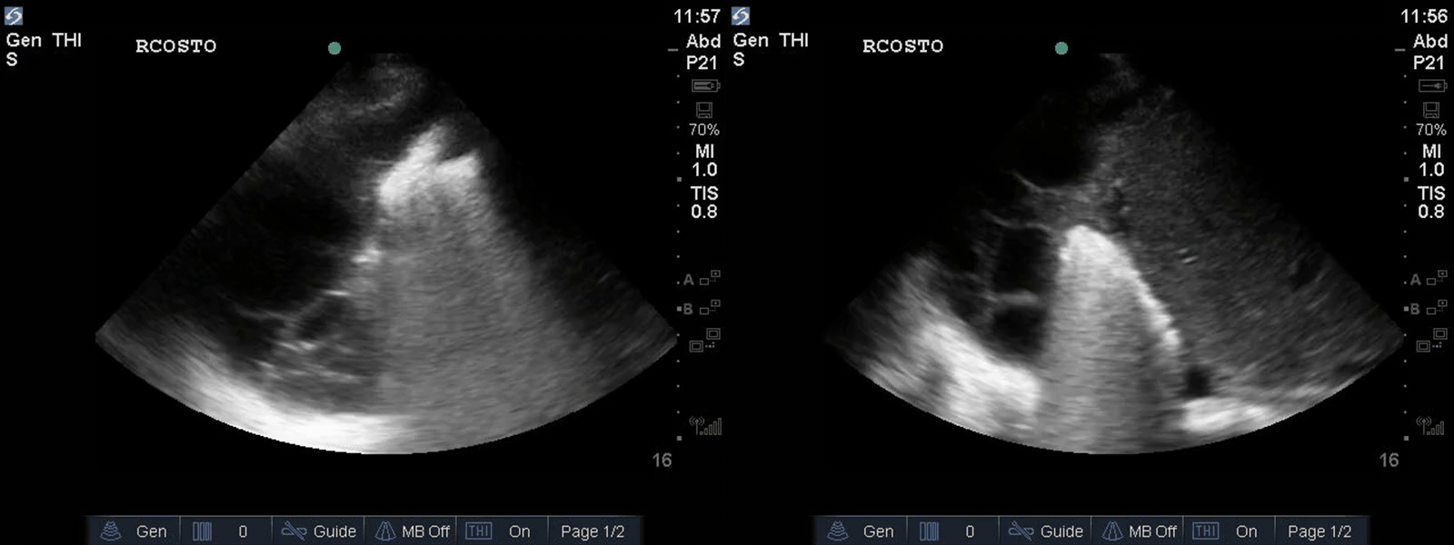
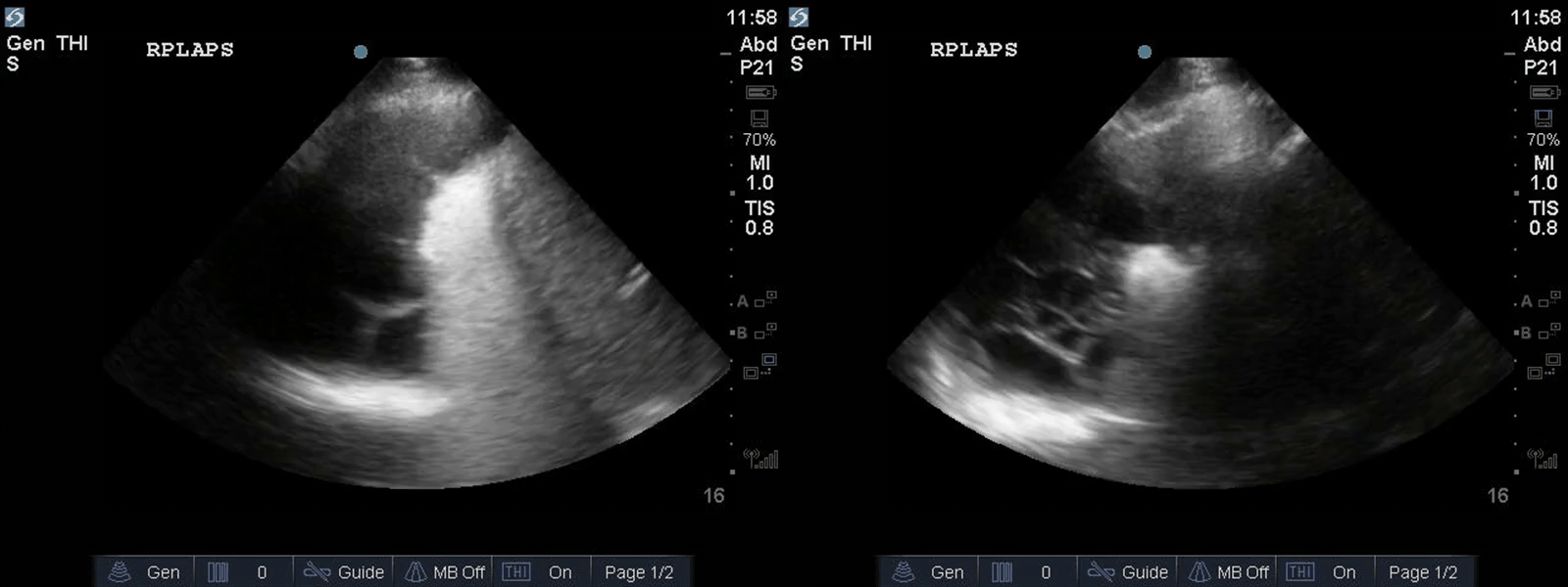
The first thing to diagnose is the presence of bilateral pleural effusions. The right appears loculated and larger than the left and you can actually see trapped aerated lung far field. All effusions should also be closely examined for complexity. Simple, free flowing, anechoic effusions are usually consistent with a transudative etiology. In contrast, echogenic material, with or without septations, represents a complex effusion and is suggestive of an exudative etiology.
This case illustrates very complex, heavily septated bilateral pleural effusions. Given the clinical context of persistent MRSA bacteremia, this is highly suggestive of bilateral empyema which should be managed with tube thoracostomy drainage. In this case as well, the sonographic evidence of extensive septations predicts the need for intrapleural fibrinolytic therapy (tPA/DNase), longer duration of drainage, and potentially, even the need for VATS decortication.
POCUS in this case was able to identify extensive, complex bilateral pleural effusions which were likely driving ongoing bacteremia and sepsis which expedited appropriate management. See the attached articles below for further learning on this!
Chen_et_al-2000-Journal_of_Ultrasound_in_Medicine
Soni – Ultrasound diagnosis Management of pleureal effusions


It’s realy look like a bilateral pleural effusion. In the right i Can see some fibrin attachement. Probably a loculated pleural effusion. It could be difficult to perform a chest tube in this situation because the needle Can go trough the lung parenchyma due to the fibrin attachement so i think it would be more safe to bring her aigain to the OR. Send a sample of pleural fluid to the lab, check for infection and malignancy, put a large spectrum antibiotics by waiting for the results. Thanks for the case.