

Case of the Week: September 12th, 2019
It’s a 78 yo M with unwitnessed syncope, a subsequent tib-fib fracture, who was eventually admitted to the ICU for persistent hypotension and altered LOC that had not been fully elucidated. He had an extensive work up including a negative CTPA, CT head, and ultimately even an angiogram (based on some transient diffuse ST depression and a positive troponin) which showed clean coronary arteries. He eventually stabilized with good supportive care, and the ICU team was now trying to wean him off the ventilator and were aggressively diuresing him. They asked for a POCUS assessment to help guide further volume management. Have a look at the images. What two major findings are most striking? Should the team continue to diurese him or perhaps give some volume back?
Case of the Week: September 6, 2019
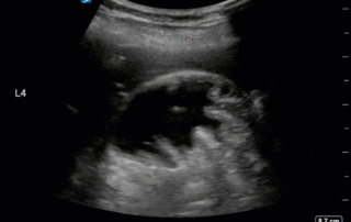
This is a 39 yo F post cardiac arrest NYD. She was in the weaning stages of her care and close to extubation, but she became newly febrile with increasing oxygen requirements. Her sputum culture was positive for E. coli. A portable CXR was done which did not show any obvious large consolidations. A POCUS thoracic study was performed. An unusual finding was seen on the left side (shown in the clips below). Also, to orient those who aren't familiar with the WesternSono shorthand here is a legend for the labels: L1 = Left anterior chest wall, L2 = Left anterior axillary line, L3 = Left costophrenic view, and L4 = Left PLAPS (PosteroLateral Alveolar and/or Pleural Syndrome)

A Decade of Ultrasound: CRUS 2019
This year marked the 10th anniverary for the annual Canadian [...]

Case of the Week: August 26, 2019
This is 54 yo M who presented for an elective surgery. On POD # 0 he became tachycardic with subjective dyspnea and hypotension progressing into a PEA arrest. ROSC was quickly obtained with typical ACLS and he was placed on life support and transferred to the ICU for further management. A CTPA was negative, and his EKG was unremarkable with no evidence of coronary ischemia. On POD # 1 he remained hemodynamically unstable, and given very poor transthoracic windows, the decision was made to perform a TEE. Have a look at the images and Doppler information below. Is there a finding that may explain the etiology of his arrest? What would your recommendation be to the treating team?

Case of the Week: August 19, 2019
This is a 35 yo M PWID who presented with a right septic AC joint, MRSA bacteremia and hypoxic respiratory failure. He was taken to the OR for washout of his AC joint and subsequently transferred to the ICU for post-op management. A post-operative CXR showed some patchy consolidations but no obvious pleural effusions. The POCUS team was subsequently deployed. Interrogation at the costophrenic angle and PLAPS (posteroalveolar and/or pleural syndrome) point on both sides yielded the following images. What do you see and what should the next steps in management be?

Case of the Week: June 6, 2019
A 47 year-old female is admitted to ICU for respiratory failure and sepsis. She has a history of immune suppression and has had a prolonged stay in ICU. She is requiring pressors and the POCUS team was asked to assess cardiac function. Here are some of her echo images: