About Derek Wu

This author has not yet filled in any details.
So far Derek Wu has created 60 blog entries.

Advanced Critical Care Ultrasound Quantitative Assessment Resource

Our previous Critical Care Ultrasound Fellow, Marko Balan MD FRCPC, who is now an intensivist at Dartmouth General Hospital has put together a comprehensive, point-of-care resource containing a wealth of advanced critical care ultrasound material. You will find in it extensive coverage of quantitative techniques that can be used for the assessment of the critically ill. Topics include: hemodynamic assessment, diastology, valvular assessment, pericardial assessment, [...]

By |2020-04-22T23:21:15+00:00April 22nd, 2020|Acquisitions, Echo, Hemodynamics, Lung Ultrasound, Resources|Comments Off on Advanced Critical Care Ultrasound Quantitative Assessment Resource

Case of the Week: February 14, 2020

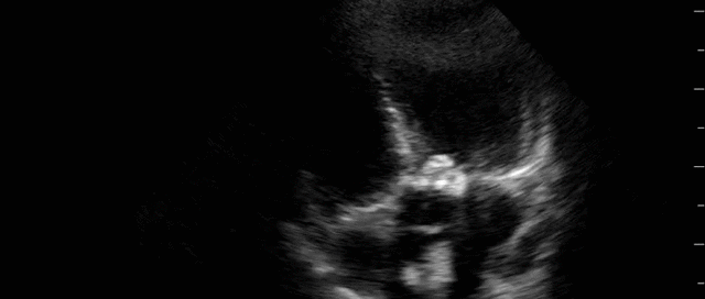
This is a 58yo F with ESRD with a history of known infective endocarditis of her AV for which she had previously completed a prolonged course of IV antibiotics. She was admitted to the ICU for worsening dyspnea. The POCUS team performed cardiac exam with some of the most striking clips shown below. What do you think? What complication of her endocarditis is shown and how would you grade its severity?

By |2020-05-28T06:49:18+00:00February 15th, 2020|Case of the Week, Cases|Comments Off on Case of the Week: February 14, 2020

Case of the Week: February 6, 2020

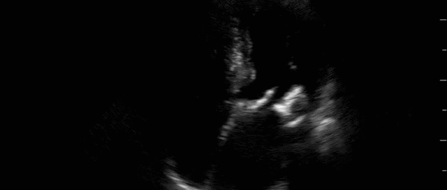
This was a case of an 80 yo M, previously healthy, with no known significant previous medical history. He presented to the ED with acute decreased level of consciousness. The leading concern was acute spontaneous ICH. He was intubated and taken for a CT Head which surprisingly was completely normal. He was subsequently transferred to the ICU with the diagnosis of altered LOC NYD. A POCUS study was performed, and several cardiac clips are shown below. What do you think? Does this give a potential clue to what could be going on?

By |2020-07-26T22:37:02+00:00February 9th, 2020|Case of the Week, Cases|2 Comments

Case of the Week: January 29, 2020

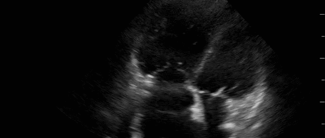
This is 65 yo M who presented with acute hypotension, hypoxia, and worsening AKI in the setting of known severe pulmonary hypertension secondary to a previous PE (CTEPH). He was admitted to the ICU for consideration of CRRT and IV flolan. A few days into his admission, the team asked for a POCUS cardiac assessment to reassess the status of his RV. Have a look the images below. Specifically, what do you think about the tricuspid regurgitation and estimated RVSP?

By |2020-07-26T22:38:02+00:00January 31st, 2020|Case of the Week, Cases|Comments Off on Case of the Week: January 29, 2020

Case of the Week: January 22, 2020

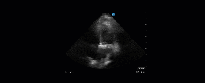
This is a 74yo F with a past medical history significant for ESRD and severe PVD who was transferred to the ICU overnight with presumed septic shock thought to be related to left foot osteomyelitis. She had presented to the ED with refractory hypotension and altered LOC necessitating intubation and high dose vasopressors. The POCUS team went to do a focused cardiac exam, primarily to see if there was a cardiogenic component to his shock. The following TTE images were taken and the decision was made to perform a point-of-care TEE. What do you think is going on?

By |2020-09-19T05:49:15+00:00January 23rd, 2020|Case of the Week, Cases|Comments Off on Case of the Week: January 22, 2020

Case of the Week: December 18, 2019

This is a 70yo F with a recent diagnosis of left sided lung cancer. She presented with hemoptysis and was subsequently admitted to the ICU for monitoring.  There she was found to be tachycardic at 120bpm and had reduced urine output. The patient had no known cardiac history and no previous echocardiogram on file. The POCUS team performed a focused echocardiogram, given that she was a newly admitted patient with ongoing tachycardia. 

By |2019-12-19T03:22:55+00:00December 19th, 2019|Case of the Week, Cases|Comments Off on Case of the Week: December 18, 2019

Case of the Week: December 9, 2019

This week's case is a 67-year old woman who presented for an outpatient EGD. Post-procedurally she became hypoxic, so was brought to the ICU for monitoring and NIPPV. She was initially hemodynamically stable; however, over the next 6 hours she became profoundly hypotensive requiring very high doses of multiple vasoactive agents. She had a PMHx significant for severe COPD and HFpEF, with an Echo from earlier this year showing an EF of 50%.Below are her the clips taken upon arrival of the POCUS team the next morning. What do you think is causing her shock based on these images? You might want to make yourself comfortable, cause this week's case is a doozy and has a lot to unpack.*Note: given her marked hyperinflation she had no obtainable traditional parasternal or apical windows, so all views were obtained from a modified subxiphoid position (hence the unconventional axis). These scans are a good lesson, however, that you can still get lots of important information even when your views aren't perfect!

By |2020-09-19T05:51:00+00:00December 9th, 2019|Case of the Week, Cases|1 Comment

Case of the Week: December 4, 2019

85 yo M polytrauma from a high-speed MVC. Now recovering in the ICU, still intubated, but over the last few days has had worsening oliguric AKI and is now on 10mcg/min of NE. The POCUS team was asked to perform a “volume status” assessment. This is one of the most common requests we get. Below is a representative look at a typical set of images we try to acquire to help answer the million-dollar question of whether a patient would benefit from fluids. Have a look at the images below (which, be forewarned, are quite Doppler heavy) and see what you think!

By |2019-12-06T22:27:22+00:00December 4th, 2019|Case of the Week, Cases|Comments Off on Case of the Week: December 4, 2019

Case of the Week: November 20, 2019

This is a case of a 52 yo F trauma patient with severe traumatic brain injury. She was intubated and transferred to the ICU where, given the severity of her TBI, an intraparenchymal ICP monitor was placed by neurosurgery. The ICP monitor was initially showing an ICP of 10. Overnight, however, despite no apparent clinical change, the ICP waveform was inconsistent and the readings were now showing values around 40-50. We thus performed a point-of-care TCD to help determine whether her ICP was truly elevated or whether the ICP monitor was giving spurious information. Have a look at the following images. Do you think the TCD spectral waveform is consistent with markedly elevated ICP?

By |2019-11-20T18:24:02+00:00November 20th, 2019|Case of the Week, Cases|Comments Off on Case of the Week: November 20, 2019
Go to Top