User Avatar

About Derek Wu

This author has not yet filled in any details.
So far Derek Wu has created 60 blog entries.

Case of the Week: November 11, 2019

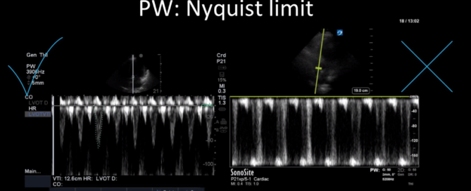
This is a 40-year old man status post-liver transplant (several months prior) who has had a complicated hospital course. He is now re-admitted with presumed septic shock (query HAP) and has been intubated for respiratory failure. He's also had a formal abdominal ultrasound commenting on periportal hepatic edema and signs of congestion.Based on the images below what interventions might you make to the team in addition to antimicrobial therapy? The team is particularly worried about right heart failure and elevated right-sided pressures - what do you tell them based on your findings?

By |2019-11-11T17:45:58+00:00November 11th, 2019|Case of the Week, Cases|Comments Off on Case of the Week: November 11, 2019

Case of the Week: November 7, 2019

This is a 54 yo M who presented with acute hypoxic respiratory failure necessitating intubation and ICU admission. Post-intubation he had a P/F ratio of 54 despite a relatively unremarkable CXR. The PEEP was increased to try to improve oxygenation. A CTPA was ordered which was negative for PE and showed just a small right lower lobe consolidation. Given that the refractory hypoxia seemed discordant with the pulmonary pathology, concern was raised for an intracardiac shunt. A point-of-care TEE was performed including a bubble study. Have a look at the selected clips below and see what you think!

By |2019-11-07T14:57:11+00:00November 7th, 2019|Case of the Week, Cases|1 Comment

Case of the Week: October 28, 2019

This is a 74-year old gentleman with multiple medical comorbidities who was admitted to the ICU with septic shock and bacteremia. He's had a tumultuous course in the unit and has been difficult to wean from vasoactive agents. Below are two series of images. The first were taken earlier in his stay, while on higher doses of vasoactive agents and more acutely unwell. The second were taken a week later when he was clinically significantly improved and weaned from inotropic support; however, there was concerned that some of his Echo parameters had actually worsened! What are we looking at here, and what's the explanation?

By |2019-10-28T09:28:50+00:00October 28th, 2019|Case of the Week, Cases|Comments Off on Case of the Week: October 28, 2019

Case of the Week: October 22, 2019

This is a case of a 56 yo F admitted with sepsis and MSSA bacteremia. A CT Head revealed several lesions concerning for septic emboli. She had known chronic significant mitral valve pathology documented prior to her admission. The POCUS team keen to see this pathology and to look for any additional signs of infective endocarditis. What do you notice about the valve on the 2D echo images and what specifically do you see on its colour doppler interrogation?

By |2020-06-07T21:13:53+00:00October 24th, 2019|Case of the Week, Cases|Comments Off on Case of the Week: October 22, 2019

Principles of Doppler

Principles of Doppler Learn the principles of Doppler ultrasound in this 20-minute screencast from Dr. Katie Wiskar. This tutorial covers basic ultrasound physics, different Doppler modes, and multiple examples from various POCUS applications.Relevant ScreencastsTutorial on aortic stenosis vs sclerosis: https://westernsono.ca/aortic-stenosis-vs-sclerosis/Tutorial on transcranial Doppler: https://westernsono.ca/screencasts/miscellaneous/trans-cranial-doppler-tcd/Tutorial on calculating stroke volume using LVOT VTI: https://westernsono.ca/screencasts/echo/stroke-volume-determination/

By |2020-04-22T22:49:38+00:00October 21st, 2019|Uncategorized|Comments Off on Principles of Doppler

Case of the Week: October 15, 2019

This is a 78-year old woman admitted a week prior with respiratory failure secondary to CHF exacerbation and COPD; on admission, she had a pleural effusion that was tapped and found to be transudative. She now has ongoing dyspnea in the ICU with increasing oxygen requirements. so the POCUS team was called in to help sort things out.

By |2019-10-21T23:30:52+00:00October 15th, 2019|Case of the Week, Cases|1 Comment

Case of the Week: October 2, 2019

81F admitted to ICU after polytrauma from an unwitnessed fall. Known history of coronary artery disease, but no previous echocardiogram on file. The POCUS team was therefore asked to assess his cardiac function. Have a look at the images below with a focus on the LV systolic function. What do you think? Is there anything in particular that stands out?

By |2019-10-03T23:05:01+00:00October 3rd, 2019|Case of the Week, Cases|Comments Off on Case of the Week: October 2, 2019

Case of the Week: September 18, 2019

This week is a case of a 29 yo F with known severe pulmonary hypertension. The etiology was thought secondary to cocaine-induced idiopathic pulmonary arterial hypertension. Unfortunately, she sustained a cardiac arrest (you’ll see why when you look at the images). ROSC was obtained and she was transferred to the ICU. Despite maximal medical support of her RV (optimal ventilator management, IV flolan, inhaled nitric oxide, inotropes including milrinone and vasopressin) she had persistent hypotension and worsening renal failure necessitating CRRT. The overnight team decided to trial some small boluses of crystalloid to see if that would help. Have a look at the images below and decide whether or not you would give fluids or recommend something else? If I said the CVP (as measured from the right IJ central line) was 22, what would the estimated RVSP be?

By |2019-09-19T01:52:52+00:00September 18th, 2019|Case of the Week, Cases|1 Comment

Case of the Week: September 12th, 2019

It’s a 78 yo M with unwitnessed syncope, a subsequent tib-fib fracture, who was eventually admitted to the ICU for persistent hypotension and altered LOC that had not been fully elucidated. He had an extensive work up including a negative CTPA, CT head, and ultimately even an angiogram (based on some transient diffuse ST depression and a positive troponin) which showed clean coronary arteries. He eventually stabilized with good supportive care, and the ICU team was now trying to wean him off the ventilator and were aggressively diuresing him. They asked for a POCUS assessment to help guide further volume management. Have a look at the images. What two major findings are most striking? Should the team continue to diurese him or perhaps give some volume back?

By |2019-09-12T16:24:42+00:00September 12th, 2019|Case of the Week, Cases|Comments Off on Case of the Week: September 12th, 2019

Case of the Week: September 6, 2019

This is a 39 yo F post cardiac arrest NYD. She was in the weaning stages of her care and close to extubation, but she became newly febrile with increasing oxygen requirements. Her sputum culture was positive for E. coli. A portable CXR was done which did not show any obvious large consolidations. A POCUS thoracic study was performed. An unusual finding was seen on the left side (shown in the clips below). Also, to orient those who aren't familiar with the WesternSono shorthand here is a legend for the labels: L1 = Left anterior chest wall, L2 = Left anterior axillary line, L3 = Left costophrenic view, and L4 = Left PLAPS (PosteroLateral Alveolar and/or Pleural Syndrome)

By |2019-09-07T02:36:01+00:00September 7th, 2019|Case of the Week, Cases|Comments Off on Case of the Week: September 6, 2019
Go to Top