User Avatar

About Derek Wu

This author has not yet filled in any details.
So far Derek Wu has created 60 blog entries.

A Decade of Ultrasound: CRUS 2019

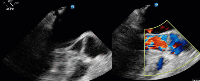
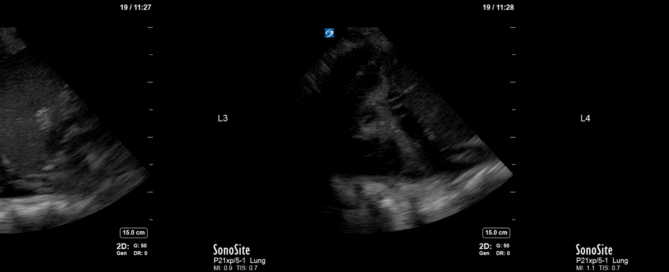
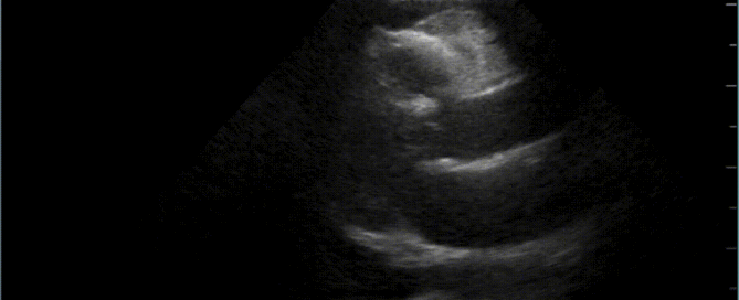
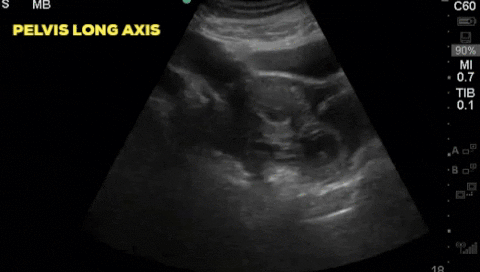
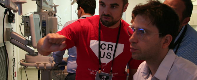
This year marked the 10th anniverary for the annual Canadian Resuscitative Ultrasound (CRUS) Course at Western University. Hosted at the sophisticated CSTAR (Canadian Surgical Technologies & Advanced Robotics) facility, the CRUS course drew trainees from across Canada to teach concepts in point-of-care ultrasound for use in the acutely ill patients typically seen in the ICU, the ED or OR. Participants engaged in didactic and hands-on [...]

By |2019-08-31T16:22:03+00:00August 30th, 2019|Courses, Critical Care Ultrasound Course, CRUS|Comments Off on A Decade of Ultrasound: CRUS 2019

Case of the Week: August 26, 2019

This is 54 yo M who presented for an elective surgery. On POD # 0 he became tachycardic with subjective dyspnea and hypotension progressing into a PEA arrest. ROSC was quickly obtained with typical ACLS and he was placed on life support and transferred to the ICU for further management. A CTPA was negative, and his EKG was unremarkable with no evidence of coronary ischemia. On POD # 1 he remained hemodynamically unstable, and given very poor transthoracic windows, the decision was made to perform a TEE. Have a look at the images and Doppler information below. Is there a finding that may explain the etiology of his arrest? What would your recommendation be to the treating team?

By |2019-08-29T02:45:17+00:00August 29th, 2019|Case of the Week, Cases|Comments Off on Case of the Week: August 26, 2019

Case of the Week: August 19, 2019

This is a 35 yo M PWID who presented with a right septic AC joint, MRSA bacteremia and hypoxic respiratory failure. He was taken to the OR for washout of his AC joint and subsequently transferred to the ICU for post-op management. A post-operative CXR showed some patchy consolidations but no obvious pleural effusions. The POCUS team was subsequently deployed. Interrogation at the costophrenic angle and PLAPS (posteroalveolar and/or pleural syndrome) point on both sides yielded the following images. What do you see and what should the next steps in management be?

By |2019-08-19T14:20:35+00:00August 19th, 2019|Case of the Week, Cases|1 Comment

Best Poster at Research Day: A-LURT’ing the People on Ultrasound

The Day Last Friday I had the amazing opportunity to present our work at the Department of Medicine Resident Research Day (take a deep breath before saying this one out loud): Acquisition and Retention of Lung Ultrasound Skills by Respiratory Therapists: a point-of-care ultrasound curriculum for respiratory therapists (and breathe). Although complimentary meals are usually enough to get hungry medical students out to [...]

By |2019-05-14T19:08:53+00:00May 13th, 2019|Education, Lung Ultrasound|1 Comment

Case of the Week: April 29, 2019

A 30-year-old female, presents to the emergency department with acute lower abdominal pain. She is 9 weeks pregnant by dates and has had no formal ultrasound prior to presentation. Her initial vitals are; HR 102, BP 116/70, RR 24, T 36.2 and SpO2 98% on RA. A bedside ultrasound is completed and demonstrates:

By |2019-04-30T02:48:52+00:00April 30th, 2019|Case of the Week, Cases|Comments Off on Case of the Week: April 29, 2019

Case of the Week: January 28, 2019

Hi POCUS enthusiasts, We set a new record with >65 participants in last week's case voting. Unfortunately this will be our last COTW/COTG until we are back in action in early April. For all those who haven't signed up for automatic distribution do it now by going to westernsono.ca and filling in your name and email address. This will be the last email reminder! Now let's jump into [...]

By |2019-01-29T15:49:26+00:00January 29th, 2019|Case of the Week, Cases|1 Comment

Case of the Week: January 21, 2019

Hi POCUS enthusiasts,Thanks again to the more than 20 participants in the online voting for last week's case! For those who have yet to sign up, just go to westernsono.ca and insert your name and email into the text box on the right side of the webpage. Then get ready to flex your POCUS muscles and post your answers/votes. Or click here to access the case directly: https://westernsono.ca/category/cases/ The [...]

By |2019-01-21T05:25:38+00:00January 21st, 2019|Case of the Week, Cases|Comments Off on Case of the Week: January 21, 2019

Case of the Week: January 14th, 2018

A 42 year old female is admitted with ARDS and a pleural POCUS is performed. Due to difficulties identifying lung sliding, M-mode was used to evaluate for pneumothorax. Based on this image, does the patient has a pneumothorax? What is the 2D ultrasound image correlate of the vertical areas as shown by the arrows?

By |2019-01-14T15:39:35+00:00January 14th, 2019|Case of the Week, Cases|Comments Off on Case of the Week: January 14th, 2018
Go to Top