Best Poster at Research Day: A-LURT’ing the People on Ultrasound
The Day Last Friday I had the amazing [...]
Case of the Week: April 29, 2019
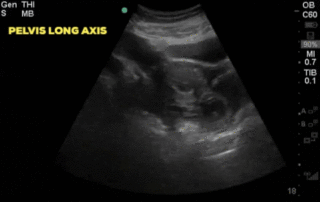
A 30-year-old female, presents to the emergency department with acute lower abdominal pain. She is 9 weeks pregnant by dates and has had no formal ultrasound prior to presentation. Her initial vitals are; HR 102, BP 116/70, RR 24, T 36.2 and SpO2 98% on RA. A bedside ultrasound is completed and demonstrates:
Case of the Week: April 17, 2019
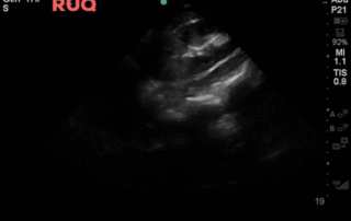
A 75 year old male presents to the emergency department with delirium and fever. You use your POCUS skills to look for a possible source of infection in his lungs.
Case of the Week: January 28, 2019
Hi POCUS enthusiasts, We set a new record with >65 participants [...]
Case of the Week: January 21, 2019
Hi POCUS enthusiasts,Thanks again to the more than 20 participants in [...]
Case of the Week: January 14th, 2018
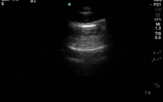
A 42 year old female is admitted with ARDS and a pleural POCUS is performed. Due to difficulties identifying lung sliding, M-mode was used to evaluate for pneumothorax. Based on this image, does the patient has a pneumothorax? What is the 2D ultrasound image correlate of the vertical areas as shown by the arrows?


