Case of the Week: October 15, 2019

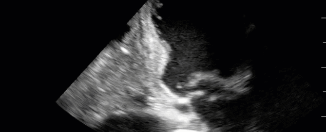
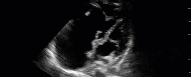
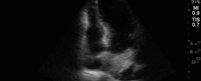
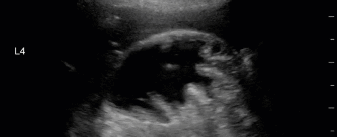
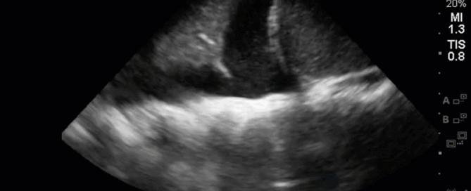
This is a 78-year old woman admitted a week prior with respiratory failure secondary to CHF exacerbation and COPD; on admission, she had a pleural effusion that was tapped and found to be transudative. She now has ongoing dyspnea in the ICU with increasing oxygen requirements. so the POCUS team was called in to help sort things out.

By |2019-10-21T23:30:52+00:00October 15th, 2019|Case of the Week, Cases|1 Comment

Case of the Week: October 2, 2019

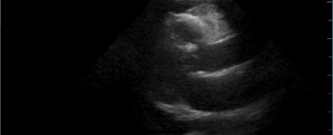
81F admitted to ICU after polytrauma from an unwitnessed fall. Known history of coronary artery disease, but no previous echocardiogram on file. The POCUS team was therefore asked to assess his cardiac function. Have a look at the images below with a focus on the LV systolic function. What do you think? Is there anything in particular that stands out?

By |2019-10-03T23:05:01+00:00October 3rd, 2019|Case of the Week, Cases|Comments Off on Case of the Week: October 2, 2019

Case of the Week: September 18, 2019

This week is a case of a 29 yo F with known severe pulmonary hypertension. The etiology was thought secondary to cocaine-induced idiopathic pulmonary arterial hypertension. Unfortunately, she sustained a cardiac arrest (you’ll see why when you look at the images). ROSC was obtained and she was transferred to the ICU. Despite maximal medical support of her RV (optimal ventilator management, IV flolan, inhaled nitric oxide, inotropes including milrinone and vasopressin) she had persistent hypotension and worsening renal failure necessitating CRRT. The overnight team decided to trial some small boluses of crystalloid to see if that would help. Have a look at the images below and decide whether or not you would give fluids or recommend something else? If I said the CVP (as measured from the right IJ central line) was 22, what would the estimated RVSP be?

By |2019-09-19T01:52:52+00:00September 18th, 2019|Case of the Week, Cases|1 Comment

Case of the Week: September 12th, 2019

It’s a 78 yo M with unwitnessed syncope, a subsequent tib-fib fracture, who was eventually admitted to the ICU for persistent hypotension and altered LOC that had not been fully elucidated. He had an extensive work up including a negative CTPA, CT head, and ultimately even an angiogram (based on some transient diffuse ST depression and a positive troponin) which showed clean coronary arteries. He eventually stabilized with good supportive care, and the ICU team was now trying to wean him off the ventilator and were aggressively diuresing him. They asked for a POCUS assessment to help guide further volume management. Have a look at the images. What two major findings are most striking? Should the team continue to diurese him or perhaps give some volume back?

By |2019-09-12T16:24:42+00:00September 12th, 2019|Case of the Week, Cases|Comments Off on Case of the Week: September 12th, 2019

Case of the Week: September 6, 2019

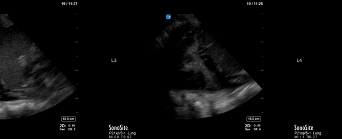
This is a 39 yo F post cardiac arrest NYD. She was in the weaning stages of her care and close to extubation, but she became newly febrile with increasing oxygen requirements. Her sputum culture was positive for E. coli. A portable CXR was done which did not show any obvious large consolidations. A POCUS thoracic study was performed. An unusual finding was seen on the left side (shown in the clips below). Also, to orient those who aren't familiar with the WesternSono shorthand here is a legend for the labels: L1 = Left anterior chest wall, L2 = Left anterior axillary line, L3 = Left costophrenic view, and L4 = Left PLAPS (PosteroLateral Alveolar and/or Pleural Syndrome)

By |2019-09-07T02:36:01+00:00September 7th, 2019|Case of the Week, Cases|Comments Off on Case of the Week: September 6, 2019

A Decade of Ultrasound: CRUS 2019

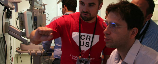
This year marked the 10th anniverary for the annual Canadian Resuscitative Ultrasound (CRUS) Course at Western University. Hosted at the sophisticated CSTAR (Canadian Surgical Technologies & Advanced Robotics) facility, the CRUS course drew trainees from across Canada to teach concepts in point-of-care ultrasound for use in the acutely ill patients typically seen in the ICU, the ED or OR. Participants engaged in didactic and hands-on [...]

By |2019-08-31T16:22:03+00:00August 30th, 2019|Courses, Critical Care Ultrasound Course, CRUS|Comments Off on A Decade of Ultrasound: CRUS 2019

Case of the Week: August 26, 2019

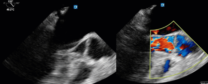
This is 54 yo M who presented for an elective surgery. On POD # 0 he became tachycardic with subjective dyspnea and hypotension progressing into a PEA arrest. ROSC was quickly obtained with typical ACLS and he was placed on life support and transferred to the ICU for further management. A CTPA was negative, and his EKG was unremarkable with no evidence of coronary ischemia. On POD # 1 he remained hemodynamically unstable, and given very poor transthoracic windows, the decision was made to perform a TEE. Have a look at the images and Doppler information below. Is there a finding that may explain the etiology of his arrest? What would your recommendation be to the treating team?

By |2019-08-29T02:45:17+00:00August 29th, 2019|Case of the Week, Cases|Comments Off on Case of the Week: August 26, 2019

Case of the Week: August 19, 2019

This is a 35 yo M PWID who presented with a right septic AC joint, MRSA bacteremia and hypoxic respiratory failure. He was taken to the OR for washout of his AC joint and subsequently transferred to the ICU for post-op management. A post-operative CXR showed some patchy consolidations but no obvious pleural effusions. The POCUS team was subsequently deployed. Interrogation at the costophrenic angle and PLAPS (posteroalveolar and/or pleural syndrome) point on both sides yielded the following images. What do you see and what should the next steps in management be?

By |2019-08-19T14:20:35+00:00August 19th, 2019|Case of the Week, Cases|1 Comment

Best Poster at Research Day: A-LURT’ing the People on Ultrasound

The Day Last Friday I had the amazing opportunity to present our work at the Department of Medicine Resident Research Day (take a deep breath before saying this one out loud): Acquisition and Retention of Lung Ultrasound Skills by Respiratory Therapists: a point-of-care ultrasound curriculum for respiratory therapists (and breathe). Although complimentary meals are usually enough to get hungry medical students out to [...]

By |2019-05-14T19:08:53+00:00May 13th, 2019|Education, Lung Ultrasound|1 Comment
Go to Top