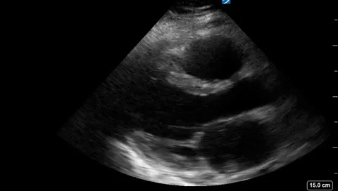
Case of the Week: April 29, 2019

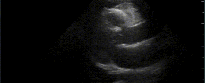
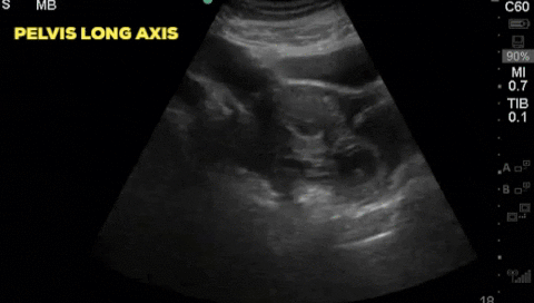
A 30-year-old female, presents to the emergency department with acute lower abdominal pain. She is 9 weeks pregnant by dates and has had no formal ultrasound prior to presentation. Her initial vitals are; HR 102, BP 116/70, RR 24, T 36.2 and SpO2 98% on RA. A bedside ultrasound is completed and demonstrates:

By |2019-04-30T02:48:52+00:00April 30th, 2019|Case of the Week, Cases|Comments Off on Case of the Week: April 29, 2019

Case of the Week: January 28, 2019

Hi POCUS enthusiasts, We set a new record with >65 participants in last week's case voting. Unfortunately this will be our last COTW/COTG until we are back in action in early April. For all those who haven't signed up for automatic distribution do it now by going to westernsono.ca and filling in your name and email address. This will be the last email reminder! Now let's jump into [...]

By |2019-01-29T15:49:26+00:00January 29th, 2019|Case of the Week, Cases|1 Comment

Case of the Week: January 21, 2019

Hi POCUS enthusiasts,Thanks again to the more than 20 participants in the online voting for last week's case! For those who have yet to sign up, just go to westernsono.ca and insert your name and email into the text box on the right side of the webpage. Then get ready to flex your POCUS muscles and post your answers/votes. Or click here to access the case directly: https://westernsono.ca/category/cases/ The [...]

By |2019-01-21T05:25:38+00:00January 21st, 2019|Case of the Week, Cases|Comments Off on Case of the Week: January 21, 2019

Case of the Week: January 14th, 2018

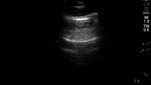
A 42 year old female is admitted with ARDS and a pleural POCUS is performed. Due to difficulties identifying lung sliding, M-mode was used to evaluate for pneumothorax. Based on this image, does the patient has a pneumothorax? What is the 2D ultrasound image correlate of the vertical areas as shown by the arrows?

By |2019-01-14T15:39:35+00:00January 14th, 2019|Case of the Week, Cases|Comments Off on Case of the Week: January 14th, 2018

Case of the Week: January 7th, 2019

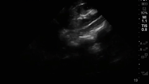
A middle aged man presented to the Emergency Department 5 days after a ERCP and stent insertion for obstructive jaundice feeling unwell, feverish, and lightheaded. He rapidly deteriorated with hypotension and was admitted to the ICU with sepsis NYD. What (if anything) is concerning on this patient's RUQ/biliary POCUS? What management steps would you recommend based on these images?

By |2019-01-07T18:57:44+00:00January 7th, 2019|Case of the Week, Cases|Comments Off on Case of the Week: January 7th, 2019

Case of the Week: December 19, 2018

Hi POCUS enthusiasts,A holiday miracle has happened (albeit a couple days late)! Kudos to Katie Wiskar for her awesome educational cases and those captivating "Eye Scandy" clips that kept us mesmerized! We can't wait to have her back. To quickly introduce myself, I'm Marko Balan and sixth-year critical care resident at Dalhousie who will be spending some time with the Western POCUS team this year. I [...]

By |2018-12-19T21:48:58+00:00December 19th, 2018|Case of the Week, Cases|1 Comment

Case of the Week: November 19, 2018

Happy Monday fellow UltraGeeks, I'm very sad to say that this is my last COTW e-mail for a while. I'll be back later in 2019 for round two as the ultrasound fellow here at Western, but for now let me say that it's been a real pleasure to share my love of ultrasound learning with you all each week. In addition, COTW will be [...]

By |2018-11-20T22:51:10+00:00November 20th, 2018|Case of the Week, Cases|Comments Off on Case of the Week: November 19, 2018
Go to Top